
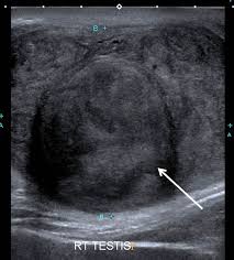
 Testis cancer is the most common cancer among adolescent and young adult males.
It typically occurs between the ages of 15 and 40 years.
There is a significant incidence of testis cancer until age 60 years, after which it is rare.
There are higher rates of TGCTs in Europe, North America, and Oceania than in Africa or Asia.
Similarly, it is more common in more-developed than less-developed regions.
Risk factors include cryptorchidism, a personal or family history of testis cancer, and HIV/AIDS.
Having a first-degree relative with testis cancer is associated with a substantially elevated risk, especially if the relative is a brother.
Male infertility is also associated with an increased risk of developing testis cancer.
Over the last century, the incidence of testis cancer has been rising substantially.
The rate of increase is slowing significantly in developed nations but not in low- and middle-income countries.
Testis cancer mortality has declined in developed countries.
The incidence to mortality ratio is 2:1 in parts of Asia and Africa vs 26:1 in Northern Europe.
Increases in incidence suggest an environmental cause, but has not been definitively identified.
The rise in testis cancer incidence has been accompanied by a rise in congenital genitourinary anomalies in baby boys as well as declining sperm counts.
No chemicals have been definitively linked to the risk of developing testis cancer.
The cell of origin for malignant GCTs is a primordial germ cell that is only present prior to birth, so that an association with pre- natal environmental exposures has been suspected for germ cell testicular cancer.
The hypothesis that prenatal or early childhood exposures may be key to determining the causes of germ cell testicular cancer.
Postpubertal males with testis cancer typically present with a testicular mass, but they may also present with testicular pain, testicular atrophy, or gynecomastia.
In patients with metastatic disease, they may present with back pain, adenopathy or thromboembolic disease
With a suspected testis a transscrotal ultrasound is performed.
If a testicle tumor is found an inguinal orchiectomy is performed to remove the involved testis and establish a histologic diagnosis.
Serum tumor markers AFP, β-HCG, and LDH should be measured before and after orchiectomy.
Imaging studies should include a computed tomography (CT) or magnetic resonance imaging (MRI) scan, both with intravenous contrast of the abdomen and pelvis, and a chest CT scan.
For seminomas, a chest x-ray is adequate for thoracic imaging if abdominal and pelvic imaging does not reveal metastatic disease.
Sperm banking is offered prior to orchiectomy if the procedure will leave them with no functioning testicle, and to all patients prior to undergoing chemotherapy, radiation therapy, or RP lymph node dissection.
3 stages of testis cancer:
Stage I GCTs are confined to the testis and epididymis
stage II GCTs include metastatic disease to the RP lymph nodes
stage III GCTs are characterized by metastatic disease beyond the RP lymph nodes.
NSGCTs with RP lymph node metastases are considered stage III rather than stage II if there are highly elevated postorchiectomy serum tumor markers (AFP >1000 ng/mL, β-HCG >5000 mU/ mL, or LDH >1.5 times the upper limit of normal).
In testis cancer, pelvic lymph node metastases are considered distant metastases because the testes’ lymphatic drainage is to the retroperitoneum.
If the markers are elevated prior to orchiectomy and return to normal afterward, then they are considered normal for staging purposes.
The biological half-life of AFP is less than 7 days.
The biological half-life of β-HCG is less than 3 days.
For patients undergoing treatment with chemotherapy, the serum tumor marker levels on day 1 of the first cycle of chemotherapy should be used for staging.
Common Chemotherapy Regimens for Germ Cell Tumors
Stage I seminoma Single-agent carboplatin
Stage I NSGCT BEP
Adjuvant therapy for patients with residual viable malignant GCT in completely resected residual masses following first-line chemotherapy VIP, TIP, or EP
Some recommend a full course of 3 cycles of BEP or 4 cycles of EP for patients with pathologic stage IIC (pN3) disease.
cBased on evidence of greater efficacy, 3 cycles of BEP is generally preferred over 4 cycles of EP for patients with good-risk disease who do not have a contraindication to bleomycin.
The tumor markers AFP, β-HCG, and LDH are important in staging, risk stratification, diagnosis, and surveillance of GCTs.
These tumor markers are not 100% sensitive nor 100% specific: roughly 25% of stage I NSGCTs and 18% of stage I seminomas relapse despite having had normal tumor markers prior to relapse, and a substantial proportion of patients have normal markers when metastatic disease is detected.
MicroRNAs are noncoding RNAs involved in gene expression regulation and can become dysregulated in patients with cancer, contributing to carcinogenesis.
For patients with GCTs, miR-371a-3p predict active germ cell malignancy with high specificity.
Serum levels of miRNA-371a-3p by polymerase chain reaction testing has a sensitivity of 90.1% and a specificity of 94% when used for the primary diagnosis of GCT, significantly out- performing conventional tumor markers.
Notably, miR- 371a-3p correlated with tumor size and stage, changed with treatment effects, and was elevated in recurrences.
Ultrasound is the preferred imaging modality for detecting testicular masses.
Staging of patients diagnosed with testis cancer should include CT scans of the abdomen and pelvis, and either a chest x-ray or CT scan of the chest.
MRI scans can be used as an alternative for abdominopelvic imaging.
Outside of assessing postchemotherapy residual masses for patients with pure seminoma, positron emission tomography (PET) scans have limited value and should not be routinely performed.
PET scans should not be used to assess treatment response for residual masses in patients with NSGCTs.
Stage I Seminoma
The prognosis for stage I seminoma is excellent, and most patients are cured by the orchiectomy alone.
Post- operatively, patients can be managed with surveillance, adjuvant radiation therapy, or adjuvant chemotherapy.
Most major guidelines recommend surveillance as the preferred management for stage I seminoma.
Multiple studies have reported 99% or higher disease-specific survival with surveillance.
Surveillance offers the benefit of reducing the risk of exposure to radiation therapy or chemotherapy, both of which are associated with acute and late toxicities.
For patients in whom surveillance is not preferable, it is recommended that single-agent carboplatin over radiation owing to studies reporting an increased mortality from secondary malignancies following radiation therapy.
A randomized controlled trial comparing a single dose of carboplatin to radiation therapy demonstrated similar relapse-free rates at 5 years (94.7% vs 96.0%) and a clear reduction in contralateral GCT for patients treated with carboplatin.
Several phase 2 studies reported consistently lower relapse rates with 2 doses of carboplatin, and we prefer 2 doses over a single dose.
Surveillance is the preferred approach for patients who are willing and able to undergo surveillance owing to the concern about the potential risk of secondary malignancies and other late toxicities from carboplatin.
Nonseminoma
For patients with clinical stage I nonseminoma and normal serum tumor markers, reasonable postoperative management options include surveillance, adjuvant chemotherapy, and nerve-sparing RP lymph node dissection (RPLND).
The presence of lymphovascular invasion (LVI) or a predominance of embryonal carcinoma histology are risk factors for relapse.
The presence of lymphovascular invasion appears to be the strongest risk factor for recurrence: One retrospective study of patients managed with active surveillance noted relapsed disease in 44% in patients with LVI vs 14% of those with- out LVI.
Trials using either 1 or 2 cycles of adjuvant bleomycin, etoposide, and cisplatin (BEP) have reduced recurrence rates to less than 5% for stage I non seminimatous teticle cancer.
RPLND can be an appropriate treatment to reduce recurrence risk while avoiding toxicity from chemotherapy.
Patients managed with surveillance who experience disease relapse will often require high- er-intensity and longer-duration treatment.
The disease-specific survival approaches 99% regardless of initial postoperative management.
Most guidelines favor surveillance for low-risk disease but differ on the preferred option for high-risk disease.
If tumor markers are persistently elevated after orchiectomy and imaging studies show no metastatic disease, the disease is classified as stage IS and should be treated similar to metastatic NSGCTs based on risk stratification.
Stage II Seminoma Stage II seminoma is generally treated with either radiation therapy or chemotherapy (BEP × 3 or EP × 4).
Historically, radiation therapy has been favored for less bulky disease (IIA and early stage IIB), although chemotherapy has been favored for bulkier disease.
Cutoffs of 3 cm and 5 cm have been used to recommend chemotherapy.
The preference of chemotherapy for men with bulkier disease is based on studies show- ing high relapse rates after radiation in such patients.
For patients with less bulky disease, chemotherapy and radiation therapy appear to have similar efficacy.
In the absence of randomized controlled trials, it is impossible to definitively recommend one modality over the other.
The relapse-free survival relapse rates were similar with radiation therapy (0%-4%) and chemotherapy (0%) in stage IIA disease, whereas the relapse rate was higher with radiation (9.5%-21.1%) than chemotherapy (0%-14.2%) in stage IIB disease.
Five-year OS ranged from 90% to 100%.
Recent studies of RPLND have reported relapse rates as high as 30%, which is substantially higher than those for radiation therapy or chemotherapy.
The benefit of reducing the risk of secondary malignancies and other late effects from those modalities may be greater than the benefit of a lower relapse rate.
Stage IIC disease (any lymph node >5 cm) should be treated with primary chemotherapy only.
Nonseminoma Stage IIA.
Treatment options for stage II NSGCT include RPLND and chemotherapy (BEP × 3 or EP × 4).
Treatment recommendations for stage II NSGCT are largely influenced by lymph node size.
If RP lymph nodes are no larger than 2 cm across at their greatest diame- ter, primary RPLND can cure the majority of patients (80%-90%) and pathologically downstage some patients to stage I if tissue shows no active GCT in the RP nodes.
RPLND gives the patient a greater likelihood of avoiding the acute and late toxicity of chemotherapy.
If RPLND reveals either pathologic stage I or IIA disease, then the standard practice is surveillance, though adjuvant chemotherapy can be considered for stage IIA disease.
The risk of relapse for stage IIA disease is only approximately 10%, and a trial comparing adjuvant chemotherapy to surveillance for pathologic stage II disease reported no difference in OS.
Adjuvant chemotherapy with 2 cycles of EP is recommended for patients with pathologic stage IIB or IIC disease because they are at much higher risk of relapse at approximately 50%.
Adjuvant chemotherapy reduces that risk to approximately 1%.
Stage IIB/C.
If nodes are more than 2 cm across at their greatest diameter, primary chemotherapy (BEP × 3 or EP × 4) is generally preferred over RPLND because the relapse rates for bulky disease are higher.
There are improved outcomes when selecting primary treatment modality of chemotherapy vs RPLND, based on risk factors, such as lymph node size.
Stage III
GCTs tend to be very sensitive to platinum-based chemotherapy, and most patients with metastatic disease can be cured.
Classification staging system should guide treatment decisions.
Good-risk disease should preferably be treated with 3 cycles of BEP.
For patients at increased risk of bleomycin pulmonary toxicity (eg, >50 years of age, chronic kidney disease, or chronic obstructive pulmonary disease or other serious lung disease) or wanting to avoid exposure to bleomycin, an alternative is 4 cycles of EP.
Similarly, carboplatin should not be substituted for cisplatin, given that carboplatin-based regimens have consistently been found to be inferior to cisplatin-based regimens.
The prognosis for good-risk disease is favorable, with a 5-year OS of approximately 90%.
Patients with good-risk disease includes those with seminoma whose metastases are limited to lymph nodes and lungs, regardless of serum tumor marker levels.
Many consider a very high β-HCG (eg, >1000 mU/mL) to be incompatible with pure seminoma, regardless of histopathologic findings, and an elevated AFP indicates that the tumor is not a pure seminoma.
An LDH level greater than 2.5 times the upper limit of normal was associated with a worse prognosis among otherwise good-risk seminoma patients (3-year progression-free survival [PFS] and OS.
Seminoma
Metastases are limited to lungs and/or lymph nodes Treatment: BEP × 3 or EP × 4
Nonseminoma Testicular or retroperitoneal primary tumor Metastases are limited to lungs and/or lymph nodes
Tumor markers: • AFP <1000 ng/mL • β-HCG <1000 mU/mL • LDH <3× ULN
Intermediate-Risk
Seminoma Presence of metastases in sites other than lungs or lymph nodes Treatment: BEP × 4 or VIP × 4 Nonseminoma (if all items present) Testicular or retroperitoneal primary tumor.
Metastases are limited to lungs and/or lymph nodes At least 1 serum tumor marker in the intermediate range and none higher than intermediate-risk range: • AFP 1000-10,000 ng/mL • β-HCG 5000-50,000 mU/mL • LDH 3-10× ULN
Poor-Risk
Nonseminoma: Mediastinal primary tumor
Presence of metastases in sites other than lungs or lymph nodes (eg, liver, brain, bones)
Tumor markers: AFP >10,000 ng/mL β-HCG >50,000 mU/mL • LDH >10× ULN
Treatment paradigms for intermediate- and poor-risk disease are similar, with risk stratification affecting prognosis rather than clinical decision-making.
The standard treatment is 4 cycles of BEP. For patients in whom bleomycin is contraindicated, etoposide, ifosfamide, and cisplatin (VIP) is an alternative regimen resulting in similar OS and PFS in both intermediate- and poor-risk disease.
Although using VIP avoids bleomycin-induced pulmonary toxicity, VIP does result in increased hematologic toxicity.
The 5-year OS for intermediate- and poor- risk disease was 79% and 48%, respectively.
More recently, data of patients with intermediate-risk seminomas had a 5-year OS of 88%, and a meta-analysis of patients with NSGCTs treated after 1989 reported pooled 5-year survival esti- mates of 83% (intermediate risk) and 71% (poor risk).
Residual masses in patients with seminoma are usually benign, and surveillance is a safe option.
The likelihood of viable residual seminoma increases with larger residual mass size.
Some advocate for surveillance of masses less than 3 cm and the use of a fluorodeoxyglucose (FDG) PET scan for masses 3 cm or larger: SEMPET trial, which reported 100% accuracy for masses larger than 3 cm.
Subsequent stud- ies have reported false-positive rates of approximately 75%, and enthusiasm for PET scans in this setting has greatly declined.
For patients with a positive PET scan, it is recommende, either repeating the PET scan at least 6 weeks later or performing resection or biopsy to confirm whether viable seminoma is present.
Enlarging residual masses on surveillance should be treated as relapsed disease and managed with chemotherapy or, less frequently, resection.
Treated seminomas are often characterized by a dense scirrhous reaction that can make resection technically difficult and increase the rate of surgical complications.
Nonseminoma If tumor markers are normal, resection of all masses larger than 1 cm is the standard of care whenever feasible.
There is no role for FDG-PET in the evaluation of residual masses in nonseminomas.
For masses in the retroperitoneum, an RPLND is performed, and masses elsewhere (eg, lungs, liver, brain) should also be resected when feasible.
Masses less than 1 cm should be observed closely.
The histopathology of residual masses may show fibrosis and necrosis, teratoma, or residual GCT.
If there is residual GCT, 2 cycles of adjuvant chemotherapy are recommended for patients who have previously received first-line but not second- or third-line chemotherapy.
A minority of patients will experience disease relapse.
For patients with stage I disease who experience relapse in the retroperitoneum, RPLND can be considered for nonbulky disease (lymph nodes <2 cm) if tumor markers are normal: of patients undergoing surveillance for stage I NSGCT who experienced disease relapse in the retroperitoneum, RPLND alone, without further therapy, was curative in 82%.
For most patients with relapsed disease, however, cytotoxic chemotherapy is the standard approach.
The choice of regimen depends on prior exposure to chemotherapy, medical comorbidities, and contraindications to specific agents.
For patients who are chemotherapy-naive or whose only prior chemotherapy was carboplatin (for stage I seminoma), chemotherapy selection should be determined by risk stratification criteria for de novo stage III disease (BEP × 3 or EP × 4, intermediate- or poor-risk: BEP × 4 or VIP × 4).
For patients who received 1 or 2 cycles of BEP for stage I or pathologic stage II NSGCT, the chemotherapy regimen used for patients at standard risk should also be given, avoiding exposing patients to more than 4 cycles of bleomycin-containing chemotherapy owing to the risk of pulmonary complications.
For patients who experience disease relapse after first- line chemotherapy for advanced-stage disease (eg, BEP × 3, EP × 4, BEP × 4, VIP × 4), treatment with either standard-dose or high-dose chemotherapy (HDCT) with autologous stem cell transplantation (ASCT) are options.
Paclitaxel, ifosfamide, and cisplatin (TIP)83 and vinblastine, ifosfamide, and cisplatin (VeIP)84 are both acceptable options for standard-dose chemotherapy, whereas carboplatin plus etoposide is the most commonly used high-dose chemotherapy regimen.
HDCT with ASCT has demonstrated efficacy in heavily pretreated patients with relapsed GCTs.
Prognosis is poor for patients with multiple disease relapses or platinum-refractory disease.
The combination of gemcitabine, oxaliplatin, and paclitaxel was shown to have efficacy in cisplatin-refractory disease or relapse after HDCT with ASCT.
The overall response was 51%, with 5% achieving a complete response.
Fifteen percent remained in remission after chemotherapy with or without residual tumor resection at a median follow-up of 5 months.
The efficacy of checkpoint inhibitors for relapsed/ refractory GCTs is disappointing.
Personalized treatment of refractory GCTs based on molecular or genomic profiling has thus far been disappointing because targetable mutations are rarely identified.
Survivorship
Radiation therapy for testis cancer has also been associated with decreased life expectancy.
Chemotherapy side effects and late toxicities include peripheral neuropathy, high-pitch hearing loss, tinnitus, cardiovascular disease, reduced pulmonary and renal function, Raynaud phenomenon, hypogonadism, and infertility.
Both radiation therapy and chemotherapy are associated with an increased risk of developing secondary malignancies, which have been associated with reduced life expectancy.
Radiation therapy and chemotherapy have also both been associated with an increased incidence of erectile dysfunction.