 A type of thoracic surgery performed using a video camera introduced into the patient’s chest via a scope.
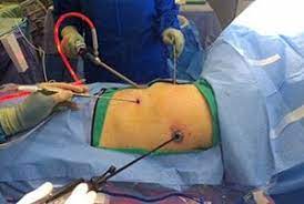
The camera and instruments are inserted through small ports reducing the risks for infection and dehiscence.
Video-assisted thoracic surgery (VATS) is a procedure to diagnose and treat certain conditions that affect the chest area.
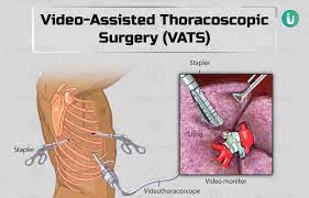
A thin tube with a tiny video camera on the end, a thoracoscope, is placed in the chest via small incision.
Video-assisted thoracoscopic surgery (VATS) is a type of minimally invasive thoracic surgery performed using a small video camera mounted to a fiberoptic thoracoscope which allows the surgeon to see inside the chest by viewing the video images relayed onto a television screen, and perform procedures using elongated surgical instruments.
Video-assisted thoracic surgery (VATS) is a procedure to diagnose and treat certain conditions that affect the chest area.
A thin tube with a tiny video camera on the end, a thoracoscope, is placed in the chest via small incision.
Video-assisted thoracoscopic surgery (VATS) is a type of minimally invasive thoracic surgery performed using a small video camera mounted to a fiberoptic thoracoscope which allows the surgeon to see inside the chest by viewing the video images relayed onto a television screen, and perform procedures using elongated surgical instruments.
The scope allows visualization of the chest cavity.
Surgical instruments are placed into the chest in separate small incisions.
The scope allows visualization of the chest cavity.
The procedure allows more rapid recovery than standard thoracic surgery using thoracotomy or sternotomy.
VATS avoids muscle splitting and bone fractures allowing for diminished duration and intensity of pain with a shorter recovery time than conventional thoracic surgery.
However, lung deflation on the side of the operated chest must occur to be able to visualize and pass instruments into the thorax.
This is usually accomplished with a double-lumen endotracheal tube that allows for single-lung ventilation, or a one-side bronchial occlusion delivered via a standard single-lumen tracheal tube.
Procedures that can be performed with VATS include: biopsy of pulmonary, pleural or mediastinal tissues, decortication, pleurodesis for recurrent pleural effusions or spontaneous pneumothorax, wedge resection of lung masses, resection of mediastinal or pleural masses, sympathectomy, operations for diaphragmatic hernias, esophageal resection, resection of esophageal masses or diverticula, lobectomy/mediastinal lymphadenectomy for lung cancer.
The instrumentation utilizes a camera-linked 5mm or 10mm fiberoptic scope, and conventional thoracic instruments or laparoscopic instruments.
Unlike laparoscopy, carbon dioxide insufflation is not generally required with VATS.
Procedure rerquires lung deflation on the side of the chest where VATS is being performed to visualize and pass instruments into the thorax.
A double-lumen endotracheal tube that allows for single lung ventilation or a bronchial blocker is utilized.
VATS also helps provide diagnoses and treat thoracic conditions like:
Lung cancer
Esophageal cancer.
Heart cancer.
Hiatal hernia.
Hyperhidrosis
Lung diseases like chronic obstructive pulmonary disease (COPD) and lung infections like tuberculosis.
Myasthenia gravis.
Pericarditis.
Pleural effusion and pleural mesothelioma.
Spine tumors and abscesses.
Thymoma and thymic carcinoma.
Thoracic surgeons use VATS to perform different procedures like:
Esophagectomy, the removal of the esophagus.
Laminectomy, the treatment of spine tumors and abscesses.
Lung volume reduction surgery (LVRS), the removal of diseased lung tissue.
Pericardiectomy, the removal of part or all of the pericardium.
Thymectomy, the removal of a diseased thymus gland.
VATS lobectomy, the removal of a lobe of a cancerous lung.
Some surgeons use robotic technology to perform video-assisted thoracoscopy.
What are the benefits of VATS?
VATS is a minimally invasive procedure.
Benefits of VATS include:
Less risk of blood loss that may require a blood transfusion.
A shorter hospital stay.
Less severe postoperative pain and scarring.
Faster recovery of respiratory function.
Lower risk of infection or complications.
A quicker return to daily activities.
VATS complications include:
Arrhythmias.
Blood clots and strokes.
Bruising.
Pneumothorax or atelectasis
Damage to nearby glands, organs, nerves or blood vessels.
Hypoxemia
Infections.
Internal bleeding and blood loss.
Respiratory problems like pneumonia.
Operations that traditionally were carried out with thoracotomy or sternotomy that today are performed with VATS include: biopsy for diagnosis of pulmonary, pleural or mediastinal pathology; decortication for empyema; pleurodesis for recurrent pleural effusions or spontaneous pneumothorax; surgical stapler-assisted wedge resection of lung masses; resection of mediastinal or pleural masses; thoracic sympathectomy for hyperhidrosis; operations for diaphragmatic hernias or paralysis; esophageal resection or resection of esophageal masses or diverticula; and VATS lobectomy/mediastinal lymphadenectomy for lung cancer.