Schwannomas are tumors that originate from Schwann cells, which are responsible for producing myelin, a substance that surrounds and insulates nerve fibers.
These tumors are usually noncancerous and develop along the peripheral nerves, often in the head, neck, or extremities.
Symptoms of schwannoma can vary depending on the location of the tumor and the nerves it affects.
Common symptoms include pain, weakness, tingling, or numbness in the affected area. If the tumor grows larger, it can potentially compress nearby structures and cause more severe symptoms.
The diagnosis of schwannoma typically involves a combination of imaging studies, such as magnetic resonance imaging (MRI), and a biopsy to confirm the presence of tumor cells.
Treatment options depend on the size and location of the schwannoma, as well as the individual’s overall health.
In some cases, the tumor may be observed to monitor its growth, while in others, surgical removal may be necessary.
A schwannoma, or neurilemmoma, i a usually benign nerve sheath tumor composed of Schwann cells, which normally produce the insulating myelin sheath covering peripheral nerves.
Schwannomas are tumors, consisting only of Schwann cells.
The tumor cells always stay on the outside of the nerve, but the tumor itself may either push the nerve aside and/or up against a bony structure.
Schwannomas are relatively slow-growing.
Less than 1% become malignant, degenerating into a form of cancer known as neurofibrosarcoma.
These masses are generally contained within a capsule, so surgical removal is often successful.
Schwannomas can be associated with neurofibromatosis type II, which may be due to a loss-of-function mutation in the protein merlin.
They are universally S-100 positive, which is a marker for cells of neural crest cell origin.
Schwannomas of the head and neck are a fairly common occurrence and are found incidentally in 3–4% of patients at autopsy.
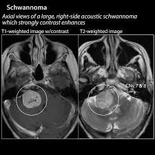
The most common are vestibular schwannoma, a tumor of the vestibulocochlear nerve that may lead to tinnitus and hearing loss on the affected side.
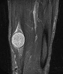
Outside the cranial nerves, schwannomas may present on the flexor surfaces of the limbs.
Rare occurrences of these tumors in the penis have been documented
Verocay bodies are seen histologically in schwannomas.
The correlation with schwannoma predisposition disorders like NF2 and schwannomatosis, is approximately 5 percent of cases.
Pleural schwannoma is extremely rare.
Schwannomas on MRI usually shows hyper or iso-intensity on T1-weighted images and heterogenous hyperintensities on T2 weighted images.
Pleural schwannoma typically shows fatty degeneration, hemorrhage, perivascular hyalinization, and cystic formation thus giving heterogenous hyperintensities on T2 weighted images.
Complete surgical removal of pleural schwannoma is the usual treatment.