Arises from the neural crest cells.
Peripheral nerves consist of sensory, motor, and autonomic fibers.
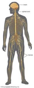
The peripheral nervous system (PNS) is one of two components that make up the nervous system the other part being the central nervous system (CNS).
The PNS consists of the nerves and ganglia outside the brain and spinal cord.
Its main function is to connect the CNS to the limbs and organs, essentially serving as a relay between the brain and spinal cord and the rest of the body.
The PNS is not protected by the vertebral column and skull, like the CNS, or by the blood–brain barrier.
As a result the PNS which leaves it exposed to toxins and mechanical injuries.
The peripheral nervous system is divided into the somatic nervous system and the autonomic nervous system.
In the somatic nervous system, the cranial nerves are part of the PNS with the exception of the optic nerve, along with the retina.
The second cranial nerve is not a true peripheral nerve but a tract of the diencephalon.
Cranial nerve ganglia originated in the CNS.
The remaining ten cranial nerve axons extend beyond the brain and are therefore considered part of the PNS.
The autonomic nervous system involuntary controls of smooth muscles and glands.
The connection between CNS and organs allows the system to be in two different functional states: sympathetic and parasympathetic.
The somatic nervous system under voluntary control.
The somatic nervous system transmits signals from the brain to end organs such as muscles.
The sensory nervous system is part of the somatic nervous system.
The sensory nervous system transmits signals from senses such as taste and touch to the spinal cord and brain.
The autonomic nervous system is a self-regulating system.
The autonomic nervous system influences the function of organs outside voluntary control, such as the heart rate, or the functions of the digestive system.
The somatic system includes the sensory nervous system and the somatosensory system and consists of sensory nerves and somatic nerves, and many nerves which hold dual functions.
In the head and neck, cranial nerves carry somatosensory data.
There are twelve cranial nerves which mainly control the functions of the anatomic structures of the head with some exceptions.
Because the nuclei of the olfactory nerve and the optic nerves lie in the forebrain and thalamus, respectively, they are not considered to be true cranial nerves.
The vagus nerve, receives sensory information from organs in the thorax and abdomen.
The accessory nerve is responsible for innervating the sternocleidomastoid and trapezius muscles, neither of which being exclusively in the head.
Sensory receptors are modified ends of sensory neurons; modified to deal with specific types of stimulus, thus there are many different types of sensory receptors in the body.
The neuron is the primary component of the nervous system, which transmits messages from sensory receptors all over the body.
Spinal nerves are responsible for somatosensory information for the rest of the body.
Spinal nerves arise from the spinal cord, usually as a web of interconnected nerves roots that arrange to form single nerves.
Spinal nerves control the functions of the rest of the body.
In humans, there are 31 pairs of spinal nerves: 8 cervical, 12 thoracic, 5 lumbar, 5 sacral, and 1 coccygeal.
Nerve roots are named according to the spinal vertebrata which they are adjacent to.
In the cervical region, the spinal nerve roots come out above the corresponding vertebrae.
From the thoracic region to the coccygeal region, the spinal nerve roots come out below the corresponding vertebrae.
In the lumbar and sacral region, the spinal nerve roots travel within the dural sac and they travel below the level of L2 as the cauda equina.
The first 4 cervical spinal nerves, C1 through C4.
C1 through C4 split and recombine to produce nerves that serve the neck and back of head.
Spinal nerve C1 is called the suboccipital nerve, which provides motor innervation to muscles at the base of the skull.
C2 and C3 form many of the nerves of the neck, providing both sensory and motor control, and include the greater occipital nerve, which provides sensation to the back of the head, the lesser occipital nerve, which provides sensation to the area behind the ears, the greater auricular nerve and the lesser auricular nerve.
The phrenic nerve is a nerve essential for our survival which arises from nerve roots C3, C4 and C5.
It supplies the thoracic diaphragm, enabling breathing. If the spinal cord is transected above C3, then spontaneous breathing is not possible.
The last four cervical spinal nerves, C5 through C8, and the first thoracic spinal nerve, T1, combine to form the brachial plexus.
The brachial plexus array of nerves, split, combine and recombine to form the nerves to the upper-limb and upper back.
Lumbosacral plexus refers to nerves from L1–coccygeal1.
The anterior divisions of the lumbar nerves, sacral nerves, and coccygeal nerve form the lumbosacral plexus.
In the lumbosacral plexus, the first lumbar nerve is frequently joined by a branch from the twelfth thoracic.
The lumbosacral plexus is usually divided into three parts:
lumbar plexus
sacral plexus
pudendal plexus
The autonomic nervous system controls involuntary responses and regulates physiological functions.
The central nervous system, brain and spinal cord, are connected with ganglionic neurons to organs that have smooth muscle, such as the heart, bladder, and other cardiac, exocrine, and endocrine related organs.
Physiological effects from autonomic activity include pupil constriction and dilation, and salivation.
The autonomic nervous system is always activated, but is either in the sympathetic or parasympathetic state.
The sympathetic or parasympathetic state can overshadow each other and result in the release of different kinds of neurotransmitters, depending on the situation.
The enteric nervous system is a division of the autonomic nervous system located only around the digestive tract.
The enteric nervous system allows for local control without input from the sympathetic or the parasympathetic branches, though it can still receive and respond to signals from the rest of the body.
The enteric system is responsible for functions related to gastrointestinal system.
The sympathetic system is activated during situations in which great mental stress or physical danger is encountered.
The sympathetic system is associated with neurotransmitters such as norepinephrine, and epinephrine.
Release of these neurotransmitters increases heart rate and blood flow in certain areas like muscle, while simultaneously decreasing activities of non-critical functions for survival, like digestion.
The parasympathetic nervous system primarily uses the neurotransmitter acetylcholine (ACh) as a mediator.
The parasympathetic system allows the body to function in a rest and digest state.
When the parasympathetic system dominates there are increases in salivation digestion activity, while heart rate and other sympathetic response decrease.
Unlike the sympathetic system, there is some voluntary controls in the parasympathetic system, such as control of urination and defecation.
Mononeuropathy refers to damage to a nerve or nerve root.
Nerve injuries can be because of trauma, or compression.
In peripheral neuropathy, the function one or more nerves are damaged.
Toxic damage to the peripheral nervous system may occur because of diabetes, alcohol, heavy metals or other toxins, some infections, autoimmune and inflammatory conditions such as amyloidosis and sarcoidosis.
Peripheral neuropathy is associated with a sensory loss in a glove and stocking distribution that begins at the periphery and slowly progresses upwards.
Peripheral neuropathy may also be associated with acute and chronic pain.
Peripheral neuropathy is not just limited to the somatosensory nerves, but the autonomic nervous system too as an autonomic neuropathy.
One reply on “Peripheral nervous system”
Hi