 A paraganglioma is a rare neuroendocrine neoplasm that may develop at various body sites: the head, neck, thorax and abdomen.
Accordingly, paragangliomas are categorized as originating from a neural cell line.
Paragangliomas originate from paraganglia in chromaffin-negative glomus cells derived from the embryonic neural crest, functioning as part of the sympathetic nervous system
These cells normally act as special chemoreceptors located along blood vessels, particularly in the carotid bodies, at the bifurcation of the common carotid artery in the neck and in aortic bodies, near the aortic arch.
When the same type of tumor is found in the adrenal gland, they are referred to as a pheochromocytoma.
They are rare tumors, with an overall estimated incidence of 1/300,000.
There is no test that determines benign from malignant tumors; long-term follow-up is therefore recommended for all individuals with paraganglioma.
Other names: chemodectoma, paraganglioma, carotid body tumor, glomus cell tumor.
Most paragangliomas are asymptomatic, present as a painless mass, or create symptoms such as hypertension, tachycardia, headache, and palpitations.
While all contain neurosecretory granules, only in 1–3% of cases is secretion of hormones such as catecholamines abundant enough to be clinically significant; in that case manifestations often resemble those of pheochromocytomas.
About 75% of paragangliomas are sporadic; the remaining 25% are hereditary.
Hereditary paragangliomas have an increased likelihood of being multiple and of developing at an earlier age.
Mutations of the genes for the succinate dehydrogenase, SDHD, SDHA, SDHC and SDHB have been identified as causing familial head and neck paragangliomas.
Mutations of SDHB play an important role in familial adrenal pheochromocytoma and extra-adrenal paraganglioma of abdomen and thorax.
Paragangliomas may also occur in MEN type 2A and 2B.
Other genes related to familial paraganglioma are SDHAF2, VHL, NF1, TMEM127, MAX and SLC25A11.
Paragangliomas appear grossly as sharply circumscribed polypoid masses and they have a firm to rubbery consistency.
They are highly vascular tumors and may have a deep red color.
Microscopically tumor cells are readily recognized as individual tumor cells that are polygonal to oval and are arranged in distinctive cell balls, called Zellballen.
Zellballen cell balls are separated by fibrovascular stroma and surrounded by sustentacular cells.
Differential diagnosis includes: related neuroendocrine tumors, such as carcinoid tumor, neuroendocrine carcinoma, and medullary carcinoma of the thyroid.
The main chief cells are located in the cell balls are positive for chromogranin, synaptophysin, neuron specific enolase, serotonin, neurofilament and Neural cell adhesion molecule; they are S-100 protein negative.
The sustentacular cells are S-100 positive and focally positive for glial fibrillary acidic protein.
Paraganglioma cells are argyrophilic, periodic acid Schiff negative, mucicarmine negative, and argentaffin negative.
About 85% of paragangliomas develop in the abdomen; only 12% develop in the chest and 3% in the head and neck region, but the latter are the most likely to be symptomatic.
Most cases are single, but rare multiple cases occur.
There are various types of head and neck paraganglioma;
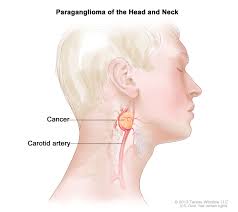
Carotid paraganglioma (carotid body tumor): Is the most common of the head and neck paragangliomas.
Carotid paraganglioma usually presents as a painless neck mass, but larger tumors may cause cranial nerve palsies, usually of the vagus nerve and hypoglossal nerve.
Glomus tympanicum and Glomus jugulare, also known as jugulotympanic paraganglioma: Both commonly present as a middle ear mass resulting in tinnitus (in 80%) and hearing loss (in 60%).
The cranial nerves of the jugular foramen may be compressed, resulting swallowing difficulty, or ipsilateral weakness of the upper trapezius and sternocleiodomastoid muscles, from compression of the spinal accessory nerve.
These patients present with a reddish bulge behind an intact ear drum:”Red drum”.
Pressure to the external ear canal with the help of a pneumatic ear speculum may make the mass blanch: Brown’s sign”.
The Organ of Zuckerkandl is a collection of paraganglia near the bifurcation of the aorta, comprising a small mass of neural crest-derived chromaffin cells, that is a common origin of abdominal paragangliomas.
Vagal paraganglioma are the least common of the head and neck paragangliomas.
Usually a gal paragangliomas present as a painless neck mass, but may result in dysphagia and hoarseness.
Pulmonary paraganglioma, occur in the lung and may be either single or multiple.
Rare sites of involvement are the larynx, nasal cavity, paranasal sinuses, thyroid gland, and the thoracic inlet, and the bladder.
Paragangliomas originate from cells of the orthosympathetic system, paragangliomas are closely related to pheochromocytomas, which however are chromaffin-positive.
Treatment:
The main treatment modalities are surgery, embolization and radiotherapy.