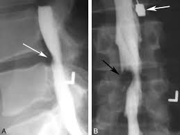
Myelography is a radiographic examination that uses a contrast medium to detect pathology of the spinal cord, including the location of a spinal cord injury, cysts, and tumors.
It involves the injection of a radiocontrast agent into the cervical or lumbar spine, followed by several X-ray projections.
Largely been replaced by the use of MRI scans, although the technique is still sometimes used under certain circumstances – though now usually in conjunction with CT.
It is used to look for the level of where spinal cord disease occurs or compression of the spinal cord at the neck region for those who are unable or unwilling to undergone MRI scan of the spine.
Lumbar myelography looks for the level of spinal cord disease such as lumbar nerve root compression, cauda equina syndrome, conus medullaris lesions, and spinal stenosis.
Lumbar puncture is done before injected contrast into the thecal sac.,
It is s dangerous to do lumbar puncture in those who has raised intracranial pressure.
AP, lateral, and oblique radiographic views of the lumbar spine are taken, and oblique view is used to examine the exiting nerve roots from cauda equina.
Thoracic myelography- lumbar puncture is done and contrast medium is injected into the puncture site, and then the head of the table is lowered.
CT myelography. Contrast media is injected into the thecal sac.
Myelography in children requires anesthesia for all children before 6 years old, and most of the children before 12 years old.
Lumbar puncture may damage the spinal cord due to possibility of tethered spinal cord syndrome or where the spinal cord is located below than the usual spinal termination level.
Lumbar puncture should be done at the lowest position as possible for such cases.
Water soluble non-ionic iodinated contrast agents are used as they cause very little complication, unlike oil-based dye that was used previously which can cause arachnoiditis.
A history of allergy to iodine is a contraindication for the use of iodinated contrast.
A CT scan is typically performed after dye has been placed with fluoroscopic guidance into a sac-like lining surrounding the spinal cord and nerves.
When the contrast has sufficiently worked its way through the spinal cord, it is followed by X-rays or a CT scan.
MRI is used for most investigations previously performed by myelography.
A CT myelogram may be useful for patients who cannot undergo MRI ; pacemakers or cochlear implants, or for those in whom MRI provides limited information (extensive metal in the spine).
Complications:
Headache occurs in about 25% of procedures and the incidence is more frequent in females.
5% of those who undergone the procedure may experience nausea and vomiting.
Risk of contrast medium being injected into the subdural space.