 A localized cutaneous form of scleroderma usually unaccompanied by the systemic features that characterize progressive systemic scleroderma.
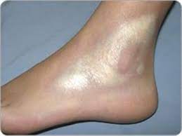
Typically present as violaceous to hypopigmented indurated plaque with a smooth, shiny surface.
Morphea most often presents as macules or plaques a few centimeters in diameter, but also may occur as bands or in guttate lesions or nodules.
Morphea is an uncommon condition that is thought to affect 2 to 4 in 100,000 people.
Most cases arise de novo.
Morphea is a thickening and hardening of the skin and subcutaneous tissues from excessive collagen deposition.
Lesions range from very small plaques only involving the skin to widespread disease causing functional and cosmetic deformities.
There is a lack lack of internal organ involvement.
There is a higher frequency of family history of autoimmune diseases in patients with morphea.
Tests for autoantibodies associated with morphea have shown results in higher frequencies of anti-histone and anti-topoisomerase IIa antibodies.
Case reports of morphea co-existing with other systemic autoimmune diseases such as primary biliary cirrhosis, vitiligo, and systemic lupus erythematosus lend support to morphea as an autoimmune disease.
Systemic manifestations include cytopenias, hypogammaglobulinemia, but scleroderma associated auto antibodies are usually not present.
Disabling pansclerotic morphia is refractory to therapy, including steroids, immuno, suppression, and autologous stem cell transplantation.
Disease pathology is attributed to abnormal collagen, synthesis and deposition. Vascular damage, abnormal immuno regulation, similar to that in other forms of scleroderma.
Borrelia burgdorferi infection may be relevant for the induction of a distinct autoimmune type of scleroderma; Borrelia-associated early onset morphea and is characterized by the combination of disease onset at younger age, infection with B. burgdorferi, and evident autoimmune phenomena as reflected by high-titer antinuclear antibodies.
Morphea–lichen sclerosus et atrophicus overlap is characterized by both lesions of morphea and lichen sclerosus et atrophicus, most commonly seen in women.
Generalized morphea is characterized by widespread indurated plaques and pigmentary changes, sometimes associated with muscle atrophy, but without visceral involvement.
Morphea profunda involves deep subcutaneous tissue, including fascia,
and tends to run a more chronic debilitating course.
Morphea profunda shows little response to corticosteroids.
Pansclerotic morphea is a form of scleroderma that involves isolated patches of hardened skin on the face, hands, and feet, or anywhere else on the body, with no internal organ involvement.
It is characterized as a systemic inflammatory process with poor wound healing with rapidly progressive, deep fibrosis, involving the mucous membranes, dermis, subcutaneous, fat, fascia, muscles, and bone leading to contractures, muscular, skeletal, atrophy, and articulate ankylosis.
Pansclerotic morphea is manifested by sclerosis of the dermis, panniculus, fascia, muscle, and at times, the bone, all causing disabling limitation of motion of joints.
Linear scleroderma is a localized scleroderma which is an autoimmune disease characterized by a line of thickened skin which can affect the bones and muscles underneath it.
Linear scleroderma most often occurs in the arms, legs, or forehead, and may occur in more than one area.
It is also most likely to be unilateral, and generally first appears in young children.
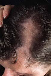
Frontal linear scleroderma is a type of linear scleroderma characterized by a linear band of atrophy and a furrow in the skin that occurs in the frontal or frontoparietal scalp.
Increased collgen production by fibroblasts with a concimitant immunogenic response with lymphocytes and plasma cells.
May be associated with radiation following breast cancer radiation.
Treatment:
Different treatments have been tried for morphea including topical, intra-lesional, systemic corticosteroids, antimalarials such as hydroxychloroquine or chloroquine have been used, immunomodulators such as methotrexate, topical tacrolimus, penicillamine, and vitamin D.
Children and teenagers with active morphea have greater improvement of disease activity or damage with oral methotrexate plus prednisone than with placebo plus prednisone.
Ultraviolet A -1 (UVA) light, with or without psoralens, is able to penetrate the deeper portions of the skin and thus, thought to soften the plaques in morphea.
Morphea is a form of scleroderma that is more common in women than men, in a ratio 3:1.
Morphea occurs in childhood as well as in adult life.