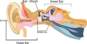
 The middle ear is the portion of the ear medial to the eardrum, and distal to the oval window of the cochlea of the inner ear.
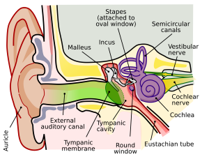
The middle ear contains three ossicles, which transfer the vibrations of the eardrum into waves in the fluid and membranes of the inner ear.
The hollow space of the middle ear is also known as the tympanic cavity and is surrounded by the tympanic part of the temporal bone.
The auditory tube, Eustachian tube , joins the tympanic cavity with the nasal cavity,nasopharynx, allowing pressure to equalize between the middle ear and throat.
The primary function of the middle ear is to transfer acoustic energy from compression waves in air to fluid–membrane waves within the cochlea.
The middle ear contains three tiny bones: ossicles, the malleus, incus, and stapes (hammer, anvil, and stirrup, respectively.)
The ossicles couple sound energy from the eardrum to the oval window of the cochlea.
The ossicles mechanically convert the vibrations of the eardrum into amplified pressure waves in the fluid of the inner ear, with a lever arm factor of 1.3.
The effective vibratory area of the eardrum is about 14 fold larger than that of the oval window, the sound pressure is concentrated, leading to a pressure gain of at least 18.1.
The eardrum is merged to the malleus, which connects to the incus, which in turn connects to the stapes.
Vibrations of the stapes footplate introduce pressure waves in the inner ear.
The lever arm ratio is variable, depending on frequency.
The ratio is generally given in relation to the tip of the malleus, the umbo, and the level of the middle of the stapes.
The eardrum is attached to the malleus handle over about a 0.5 cm distance.
The eardrum itself moves in a very chaotic fashion at frequencies >3 kHz., and the linear attachment of the eardrum to the malleus actually smooths out this chaotic motion and allows the ear to respond linearly over a wider frequency range than a point attachment.
The auditory ossicles can reduce sound pressure as the inner ear is very sensitive to overstimulation, by uncoupling each other through particular muscles.
The middle ear’s efficiency peaks at a frequency of around 1 kHz.
The movement of the ossicles may be altered/stiffened by two muscles:
The stapedius muscle, and the tensor tympani muscle.
The smallest skeletal muscle in the body is the stapedius.
The stapedius connects to the stapes and is controlled by the facial nerve.
The tensor tympani muscle is attached to the upper end of the medial surface of the handle of malleus.
The tensor tympani muscle is under the control of the medial pterygoid nerve which is a branch of the mandibular nerve of the trigeminal nerve.
The ossicle muscles contract in response to loud sounds, thereby reducing the transmission of sound to the inner ear: the acoustic reflex.
Two branches of the facial nerve that pass through the middle ear space:
the horizontal portion of the facial nerve and the chorda tympani.
Damage to the horizontal branch during ear surgery can lead to paralysis of the face.
The chorda tympani is the branch of the facial nerve that carries taste from the ipsilateral half of the tongue.
The middle ear allows the impedance matching of sound traveling in air to acoustic waves traveling in a system of fluids and membranes in the inner ear.
The middle ear couples sound from air to the fluid via the oval window, using the lever principle.
The vibratory portion of the tympanic membrane is many times the surface area of the footplate of the stapes which attaches to the oval window.
The shape of the articulated ossicular chain is a complex lever, the long arm being the long process of the malleus, the fulcrum being the body of the incus, and the short arm being the lenticular process of the incus.
The pressure of sound vibration that strikes the tympanic membrane is concentrated down to this much smaller area of the footplate, increasing the force but reducing the velocity and displacement, and thereby coupling the acoustic energy.
The middle ear can dampen sound conduction substantially when faced with very loud sound, by noise-induced reflex contraction of the middle-ear muscles.
In a high-altitude environment or on diving into water, there will be a pressure difference between the middle ear and the outside environment: the Eustachian tubes that connect the middle ear to the nasopharynx is to help keep middle ear pressure the same as air pressure.
The Eustachian tubes are normally narrowed off at the nose end, to prevent being clogged with mucus.
The Eustachian tubes may be opened by lowering and protruding the jaw; yawning or chewing helps relieve the pressure felt in the ears when on board an aircraft.
Otitis media is an inflammation of the middle ear.
The middle ear is well protected from most minor external injuries by its internal location, but is vulnerable to pressure injury, barotrauma.
Middle ear mucosa could be subjected to human papillomavirus infection, with DNAs belonging to oncogenic HV16 and HPV18, have detected in normal middle ear specimens.