 A focal lung pneumatosis, is an enclosed pocket of air or gas in the lung and includes blebs, bullae, pulmonary cysts, and lung cavities.
Blebs and bullae are classified by their wall thickness.
A bleb has a wall thickness of less than 1 mm: By radiology definition, it is up to 1 cm in total size.
A bleb originates in the pleurae, rather than in the lung parenchyma.
A bulla has a wall thickness of less than 1 mm.
A lung cyst has a wall thickness of up to 4 mm.
Cysts are walled spherical parenchymal lesions that have a well-defined interface with normal lung.
Lung cyst formation may involve ischemia, airway obstruction with distal hyperinflation, or remodeling induced matrix degrading enzymes.
A cavity has a wall thickness of more than 4 mm.
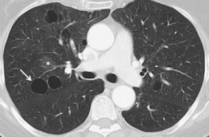
High resolution CT imaging illuminates the distribution and characteristics of cysts.
Lung cysts are seen in about 8% of the general population, with an increased prevalence in older people.
Lung cysts are not associated with emphysema.
Lung cysts may be part of the aging changes of the lungs, and cause a slight decrease in their diffusing capacity.
The presence of multiple pulmonary cysts may indicate a need to evaluate the possibility of bullous or cystic lung diseases.
Cavitation indicates workup for serious infection or lung cancer.
The most common disease causing blebs or bullae is paraseptal emphysema.
Centrilobular emphysema may sometimes be involved.
Other conditions associated with lung bullae are:
Alpha 1-antitrypsin deficiency
Marfan syndrome
Ehlers–Danlos syndromes
Cocaine smoking
Sarcoidosis
HIV/AIDS
Intravenous substance abuse with cocaine.
The cyst for example in pneumocystis pneumonia is not the same as the pulmonary cyst.
Cystic lung diseases include:
Langerhans cell histiocytosis (LCH)
Lymphangioleiomyomatosis (LAM)
Lymphocytic interstitial pneumonia
Birt–Hogg–Dubé syndrome
Pneumocystis pneumonia
Pulmonary amyloidosis
Light chain deposition disease
Lung metastases rarely cause multiple cystic lung lesions.
A focal lung pneumatosis that is an incidental imaging finding, without other suspicious findings generally does not require further follow-up.
Infections occasionally produce lung cysts.
Infections occasionally produce lung cysts.
Pneumocystis jirovecii pneumonia can cause cysts, often with lower lobe predominance.
Two infectious diseases that are commonly associated with cavities of lung tissue are Mycobacterium tuberculosis and Klebsiella pneumoniae. S
Infection with Coccidioides may result in cystic lung disease.
The formation of cavities is due to tissue necrosis and creates an environment that allows the pathogen to expand in numbers and spread further.
In the absence of infectious symptoms, a lung nodule with cavitation is a suspected lung cancer.