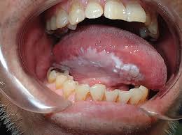
 Oral leukoplakia refers to a white plaque of variable cancer risk.
White patches of the oral cavity have potential to develop into squamous-cell carcinoma, and when this occurs, the odds surviving more than five years are poor.
Affects up to 5% of the global population, but only a small proportion of leukoplakia lesions undergo malignant transformation.
The degree of epithelial dysplasia, the lesion size, and tobacco exposure history influence transformation rates.
Between 5 and 15 percent of oral white patches are classified as dysplasia, and of these,15-20% develop into carcinoma.
Patients with normal diploid DNA content can undergo watchful waiting while patients with aneuploidy in their DNA should be treated as true carcinoma.
The term leukoplakia describes a white patch or plaque that cannot be characterized clinically or pathologically as any other disease.
Remains a clinical diagnosis of exclusion.
Leukoplakia occurs most often in middle-aged and older men and arises most frequently on the buccal mucosa, alveolar mucosa, and lower lip.
Proliferative verrucous leukoplakia is an aggressive subtype with a malignant transformation rate exceeding 10% per year, and is characterized by heterogeneous or verrucous lesions involving multiple oral subsites.
Lesions arising on the floor of mouth, lateral tongue, and lower lip are the most likely to harbor dysplasia or progress to malignancy.
The rate of progression to malignancy has been reported to be between 3.6% and 17.5%, and as many as 19.9% of leukoplakic lesions may demonstrate some degree of dysplasia, with 3.1% showing frank carcinoma.
Risk factors for malignant transformation of oral leukoplakia include advanced age, female sex, lesions of more than 200 mm2, nonhomogeneous lesions, and higher-grade dysplasia.
Immune checkpoint inhibitors may be considered for high-risk, oral pre-cancerous disease.