 Lamina cribrosa sclerae
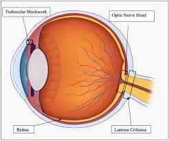
The nerve fibers forming the optic nerve exit the eye posteriorly through a hole in the sclera that is occupied by a mesh-like structure called the lamina cribrosa.
It is formed by a multilayered network of collagen fibers that insert into the scleral canal wall.
The nerve fibers of the optic nerve run through pores formed by these collagen beams.
A central retinal artery is located slightly off-center in nasal direction.
The lamina cribrosa is thought to help support the retinal ganglion cell axons as they traverse the scleral canal.
The LC is structurally weaker than the much thicker and denser sclera.
The LC is more sensitive to changes in the intraocular pressure and tends to react to increased pressure through posterior displacement.
Displacement of the lamina cribrosa causes the pores to deform and pinch the traversing nerve fibers and blood vessels causing of nerve damage in glaucoma.