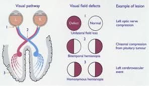
 Homonymous hemianopsia (HH) (or homonymous hemianopia) is hemianopic visual field loss on the same side of both eyes.
It is a visual field loss on the left or right side of the vertical midline.
It can affect one eye but usually affects both eyes.
HH occurs because the right half of the brain has visual pathways for the left hemifield of both eyes, and the left half of the brain has visual pathways for the right hemifield of both eyes.
When one of these pathways is damaged, the corresponding visual field is lost.
HH associated s ymptoms: clumsiness, decreased night visions, difficulty of strait line walking, distorted sight, doubled vision, turning of the head away from the side where it is present, visual hallucinations.
HH causes: brain bleed, brain inflammation, brain tumor, dementia, epilepsy, lymphoma, other kinds of brain injuries, surgery and stroke.
Homonymous hemianopsia can be congenital,but is usually caused by brain injury such as from stroke, trauma, tumors, infection, or following surgery.
Vascular and malignant or benign tumors lesions from the optic tract, to visual cortex can cause a contralateral homonymous hemianopsia.
Injury to the right side of the brain will affect the left visual fields of each eye.
Diagnostic method utilizes magnetic resonance imaging.
Mobility can be impaired for people with homonymous hemianopsia: bumping into obstacles on the side of the field loss.
A related phenomenon is hemispatial neglect, the possible neglect of the right or left: this is distinct from hemianopsia.
The more posterior the cerebral lesion is, the more symmetric or congruous the homonymous hemianopsia will be.
A transient homonymous hemianopsia can be due to a stroke, or the aura phase of migraine.
Computed tomography (CT scan) or MRI is used to investigate if stroke, tumor, structural lesion, or demyelination is the cause of homonymous hemianopsia.
Homonymous hemianopsia secondary to posterior cerebral artery occlusion – may result in memory impairment, homonymous hemianopsia, and sometimes hemisensory deficits.
The posterior cerebral artery supplies the occipital lobe and the medial portion of the temporal lobe.
Infarction of occipital cortex typically causes macular sparing hemianopias due to dual blood supply from both posterior cerebral artery and middle cerebral artery.
Prisms that bend light are prescribed for patients with hemianopsia, expanding the visual field of patients with hemifield visual defects and have the potential to improve visual function and mobility.