 Represents 10-20% of herpes zoster cases.
Frequency- Up to 125,000 per year in US.
HZO is the second most common manifestation of shingles, the first being involvement of skin.
Shingles affects up to one half million people in the United States per year, of which 10% to 25% is HZO.
Herpes zoster ophthalmicus (HZO), also known as ophthalmic zoster, is shingles involving the eye or the surrounding area.
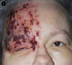
Common signs include a rash of the forehead with swelling of the eyelid.
There may also be eye pain and redness, inflammation of the conjunctiva, cornea or uvea, and sensitivity to light.
Findings include epithelial: punctate epithelial erosions and pseudodendrite.
Eye findings onset 2 to 3 days after the onset of the rash, resolving within 2–3 weeks.
Clinical findings:
Common: Nummular keratitis with anterior stromal granular deposits, occurring within 10 days of onset of rash.
Uncommon: necrotising interstitial keratitis: Characterised by stromal infiltrates, corneal thinning and possibly perforation, occurring between 3 months and several years after the onset of rash.
Rare: Disciform Keratitis, is a disc of corneal edema, folds in Descemet’s membrane, mild inflammation evident within the anterior chamber.
Chronic clinical findings.
Uncommon.
Neurotrophic: corneal nerve damage causes a persistent epithelial defect, thinning and even perforation.
Cornea susceptibility to bacterial and fungal keratitis.
Mucous plaques: linear grey elevations loosely adherent to the underlying diseased epithelium/stroma.
Anterior uveitis develops in 40–50% of people with HZO within 2 weeks of the onset of the skin rashes.
This non-granulomatous iridocyclitis is associated with:
HZO uveitis is associated with complications such as iris atrophy and secondary glaucoma are not uncommon.
Cataracts may develop in the late stages of the disease.
Fever and tingling of the skin and allodynia near the eye may precede the rash. Hutchinson’s sign: cutaneous involvement of the tip of the nose, indicating nasociliary nerve involvement.
The presence of a Hutchinson’s sign increases the likelihood of ocular complications associated with HZO.
Occurs when human herpesvirus type three reactivation presents in the first division of the trigeminal nerve, also known as the ophthalmic division.
HZO is due to reactivation of VZV within the trigeminal ganglion.
The trigeminal ganglion give rise to the three divisions of cranial nerve V (CN V), namely the ophthalmic nerve, the maxillary nerve, and the mandibular nerve.
VZV reactivation in trigeminal ganglion predominantly affects the ophthalmic nerve.
The ophthalmic nerve gives rise to three branches: the supraorbital nerve, the supratrochlear nerve, and the nasociliary nerve.
Any of these nerves can be affected in HZO, although the most feared complications occur with nasociliary nerve involvement, due to its innervation of the eye.
The supraorbital and supratrochlear nerves mainly innervate the skin of the forehead.
Complications may include visual impairment, increased pressure within the eye, chronic pain, and stroke.
It is caused by reactivation of varicella zoster virus.
HZO is due to reactivation of VZV within the trigeminal ganglion.
The underlying mechanism involves a reactivation of the latent varicella zoster virus (VZV) within the trigeminal ganglion supplying the ophthalmic nerve.
The frontal nerve is more commonly affected than the nasociliary nerve or lacrimal nerve.
Risk factors:
Poor immune function, psychological stress, and older age.
50-72% of patients demonstrate involvement of the eye itself.
Reactivation manifests with pain and perioculocutaneous rash limited to periorbital region.
Eye involvement can range from a cutaneous reaction limited to the eyelids, to corneal ulceration, to retinal disease, which may result in permanent loss of vision.
Diagnosis based on signs and symptoms.
Herpes zoster ophthalmicus as seen after fluorescence staining using cobalt blue light.
Also, fluid collected from the rash may be analyzed for VZV DNA using PCR.
Complications include: pronounced eyelid edema or skin lesions around the eye, the cornea may be affected, glaucoma, retinal necrosis, and blindness, as well as increased risk of stroke may occur.
Incidence of approximately 3.2 cases per 1000 person-years.
HZO increased incidence from 9.4 cases per 100,000 population in 2004 to 30.1 cases per hundred thousand in 2016.
The sex distribution is approximately 60% female and 40% male.
Peak incidence between ages of 50 and 79 years, with a trend towards older individuals.
Women are more likely than men to develop herpes zoster ophthalmicus as are white adults compared with black, Asian, and Latino adults.
The highest incidence rates are observed among patients over 80 years of age, at 10.9 cases 1000 person-years.
Diagnosis based on signs and symptoms.
Also, fluid collected from the rash may be analyzed for VZV DNA using PCR.
Prevention-The herpes zoster vaccine is recommended for prevention in those over the age of 50.
TREATMENT:
Treatment is generally with antiviral agents such as acyclovir, steroid eye drops and drops to dilate the pupil may also be used.
Treatment is usually with antivirals such as acyclovir, valacyclovir, or famcyclovir by mouth.
Antiviral eye drops have not been found to be useful.
Cycloplegics prevent synechiae from forming.