 Eyes are organs of the visual system
They provide the ability to receive and process visual detail, as well as enabling several photo response functions that are independent of vision.
Eyes detect light and convert it into electro-chemical impulses in neurons.
It is a complex optical system which collects light from the surrounding environment, regulates its intensity through a diaphragm, focuses it through an adjustable assembly of lenses to form an image, converts this image into a set of electrical signals, and transmits these signals to the brain through complex neural pathways that connect the eye via the optic nerve to the visual cortex and other areas of the brain.
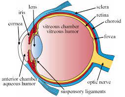
The lens and cornea, the anterior eye, focus incoming rays of light onto the retina.
The afferent visual system begins with the tear film and cornea and then encompasses the anterior chamber, crystalline lens, vitreous humor, retina, optic nerve, optic chiasm, optic tract lateral geniculate nucleus of the thalamus, optic radiation, and striate cortex in the occipital lobe, as well as specialized extrastriate areas in the parietal and temporal lobes.
The cornea, which covers the front of the eye, is the first place light enters the eye.
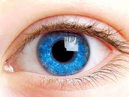
The iris is the pigmented part of the eye that determines individual eye color.
The iris dilates and contracts the pupil, regulating the amount of light that enters the eye.
The lens focuses the light onto the retina.
Rods and cones are cells in the retina that transmit light via electrical signaling to the brain, by way of the optic nerve.
Cones are responsible for color vision and sight in bright light.
Rods are helpful for sight in low light.
Refraction, refers to the bending of incoming light rays, and is primarily achieved by the cornea at the interface between air and the tear film.
Two-thirds of the 60 D refraction occurs at the cornea.
External wall made up of white tissue, the sclera, which is covered by the conjunctiva.
The front of the eye wall consists of the cornea, which is transparent.
Light enters via the cornea and passes through the pupil, the opening formed by the colored part of the eye, the iris.
The iris contracts and expands and controls the size of the pupil and therefore the amount of light that enters the eye.
The lens is behind the pupil and iris and functions to focus light entering the eye onto the retina.
The retina lines the inside of the eye and contains rods and cones, the photoreceptor cells.
The photoreceptor cells are stimulated by light focused on the retina.
The retina transmits light signals to the optic nerve, which in turn, conveys information to the brain where a visual image is perceived.
The retinal contains two types of light sensing neurons: rod photoreceptors and cone receptors.
Rod receptors respond to dim light, while cone photoreceptors respond to bright light.
Vision begins with light absorption of light by visual pigments in these photoreceptors.
Cone photoreceptors carry one of three different pigments which are maximally sensitive at long, middle, and short wave lengths, or red, green and blue, respectively.
Individuals with all three pigments are referred to as trichromats.
The absorption spectrum of the three pigments cover the visible spectrum..