 Easy, inexpensive, nonspecific test that has been used to detect conditions associated with acute and chronic inflammation, including infections, cancers, and autoimmune diseases.
The erythrocyte sedimentation rate (ESR), also called a sedimentation rate or Westergren ESR.
Refers to the rate at which red blood cells sediment in a period of one hour.
Represents the distance in millimeters that red blood cells descend in a tube over 1 hour.
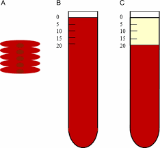
Anticoagulated blood is placed in an upright tube, the Westergren tube and the rate at which the red blood cells fall is measured and reported in mm/h.
The rate is governed by the balance between pro-sedimentation factors, mainly fibrinogen, and those factors resisting sedimentation, namely the negative charge of the erythrocytes known as the zeta potential.
The ESR being largely dependent on the elevation of fibrinogen, an acute phase reactant with a half-life of approximately one week.
Red blood cells have a slightly negative surface electrical charge, which causes them to repel each other, impeding erythrocyte package and sedimentation.
If plasma is enriched with positively charged proteins such as immunoglobulins or fibrinogen, this electrical repulsion is blunted and agglutination of red blood cells occur, speeding sedimentation.
Is considered a measure of inflammation from infection, malignancy, or rheumatologic disease, yet multiple non-inflammatory factors affect the RBC sedimentation.
Fibrinogen is a large positively charged protein and the most abundant acute phase reactant and its elevation is the predominant reason for high ESR inflammatory states.
Factors that alter the ESR include spatial interference, electrical charge, and viscosity.
Increased plasma viscosity, such as in cases of hypergammaglobuineia, can slow erythrocyte sedimentation through the plasma and decrease the ESR.
Increased spatial interference, defined as physical factors interfering with the ability of RBCs to settle in the Westergren tube, slows the packing of erythrocytes at the tube bottom, thereby decreasing the ESR.
Extreme leukocytosis impedes the RBC downward flow, which decreases the ESR.
Decreased spatial interference, such as with cases of anemia and consequently fewer RBC’s, causes an increase in the ESR because RBC’s can sediment with less obstruction.
With inflammation fibrinogen levels increase in the blood causing red blood cells to stick to each other, forming stacks called rouleaux, that tend to settle faster.
Inflammation causes cells to form dense clumps, which descend more rapidly than individual cells.
Moderate elevations may occur without a known cause, but extreme elevations of 100 mm per hour or greater are typically associated with malignancy, infection, or inflammation.
ESR is nonspecific.
Can be affected by other conditions besides inflammation.
Extreme elevations have a positive predictive value of 90% when correlated with certain diagnoses.
Extreme elevations indicator of poor prognosis.
96% of patients with extreme elevations in ESR have known diagnoses.
Diagnosis have been categorized into: infection, autoimmune, malignancy, renal disease, or miscellaneous.
Used in conjunction with other tests.
ESR is helpful in diagnosing two specific inflammatory diseases, temporal arteritis and polymyalgia rheumatica.
Also used to monitor disease activity and response to therapy in both of these diseases.
Ordered when a condition or disease is suspected of causing inflammation somewhere in the body.
There are numerous inflammatory conditions that may be detected using this test.
Moderately elevated ESR occurs with inflammation, but also with anemia, infection, pregnancy, and old age.
Normal ESR ranges increase with age.
Women have a higher normal ESR range than men.
Infection is the most common association with extreme ESR elevations.
A very high ESR usually has an obvious cause, and patients with multiple myeloma or Waldenstrom’s macroglobulinemia typically have very high levels even if they don’t have inflammation.
With polymyalgia rheumatica or temporal arteritis patients may have very high ESRs, and a rising ESR can mean an increase in inflammation or a poor response to a therapy; a decreasing ESR can mean a good response.
A low ESR is not usually a cause for concern, andvcan be seen with conditions that inhibit the normal sedimentation of RBCs, such as polycythemia, extreme leukocytosis, spherocytosis, CHF, and in sickle cell anemia.
Females tend to have a higher ESR.
Values increased in blacks.
Increased in anemias.
Menstruation and pregnancy can cause temporary elevations.
Can be used for the diagnosis and monitoring of children with rheumatoid arthritis or Kawasaki disease.
Drugs such as dextran, methyldopa, oral contraceptives, penicillamine procainamide, theophylline, and vitamin A can increase ESR
Aspirin, cortisone, and quinine may decrease it.
There is a commercial rapid test available that performs the ESR in 4 minutes by a centrifugal method.