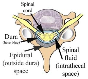
 The epidural space is a fluid-filled “potential space” in the spine located just outside the protective dural sac that houses the spinal cord.
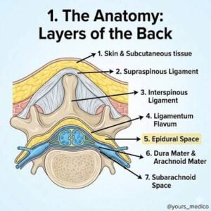
The epidural space is the anatomical space between the dura mater and the inner wall of the vertebral canal; in the spine, it contains fat, venous plexuses, lymphatics, and spinal nerve roots.
The epidural space contains loose fat, lymphatics, nerve roots, and an extensive network of blood vessels.
It spans from the base of the skull down to the sacrum.
Because of its proximity to the spinal nerves, the epidural space is heavily utilized in medicine for targeted pain management.
Local anesthetics are injected to block nerve signals, commonly used during childbirth (epidural blocks) or as regional anesthesia for surgeries.
Chronic Pain Relief: Corticosteroids can be injected into the space to dramatically reduce inflammation and swelling around compressed or irritated nerves.
Anatomical Boundaries
Anterior, The posterior longitudinal ligament and the back of the vertebral bodies.
Posterior: The vertebral laminae and the ligamentum flavum.
Lateral: The vertebral pedicles and intervertebral foramina (the openings where nerve roots exit).
It is the target site for epidural anesthesia and analgesia, where medication is placed outside the dura to affect spinal nerve roots without entering the CSF.
The spinal epidural space extends from the foramen magnum to the sacral hiatus, and its width varies by level, being narrowest in the cervical region and wider in the lumbar area.
The term can also refer to a potential space in the cranium between the skull and the outer layer of dura, but that cranial epidural space is usually absent unless pathology creates it, such as an epidural hematoma.