 Represents 1.5-11.7% of pediatric brain tumors.
Annual incidence in U.S. 0.18 per 100,000 population per year.
There are fewer than 600 papillary craniopharyngiomas per year in the US.
Onset in childhood usually between 5-14 years.
The tumors are most common in children 5 to 14 years of age and adults older than 50 years.
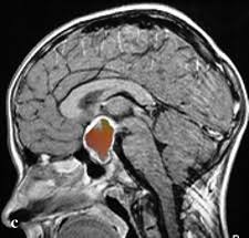
Occur in the center of the brain near the optic nerve, hypothalamus, and pituitary gland.
They grow from embryonic cells near the stalk of the pituitary gland.
A locally, aggressive neoplasm located at the midline, in or above the sella turcica
It grows adjacent to the optic chiasm, and often extends to displace the hypothalamus, pituitary, gland, third ventricle, brainstem, and major cerebral arteries.
Because of their location surgical excision is rarely feasible.
Radiation therapy is associated with short and long-term complications and variable efficacy.
uyu The natural history of these lesions includes frequent recurrences associated with slow erosion of neurologic and endocrine health.
Cause significant neurological sequelae by compressing above structures.
Tend to adhere to adjacent tissues.
Histologically benign.
2 subtypes reflect tumor location: The adamantinomatous form affects children and a papillary form that occurs primarily in adults.
Both subtypes have a high rate of gene mutations.
Childhood adamantinomatous types have a 96% mutation rate of beta-catenin gene (CTNNB1), which affects cells to cell adhesion through the Wnt pathway.
95% of adult papillary tumors have a mutation in the BRAF gene, affecting cell growth rates.
Surgery treatment of choice but cure is difficult.
Recurrence rate between 5-50%.
After subtotal resections up to 70% develop progressive disease.
Combination of surgery and radiation therapy progression of disease occurs in up to 20% of patients at 10 years and 46% the 20 years.
Overall long-term survival 80%.
Overall survival rate is 90%, but recurrence and complications from surgery and radiation can lead to impaired quality of life including: visual defects, epilepsy, hypo pituituitarism, diabetes insipidus, hypothalamic obesity, and other neurologic problems.
Lesions related to internal carotid artery involvement, anterior cerebral arteries, pituitary gland, hypothalamus, optic nerves, optic chiasm and optic tracts.
Because the tumor tends to wrap itself around vital structures of the brain standard therapy is difficult and itself can be debilitating.
Surgical and radiation therapy may be associated with visual loss, endocrine dysfunction, hypothalamic impairment, cerebral infarction impaired cognition, seizures and moyamoya syndrome.
Single or multiple cysts seen characteristically and can account for 80-90% of bulk of the tumor and 69% of patients have mainly cystic lesions.
BRAF inhibitors may be beneficial for papillary craniopharyngiomas.
Combined therapy with dabrafenib and trametinib have significant clinical response rate.
In a single group study involving patients with papillary craniopharyngioma, 15 of 16 patients had a partial response, or better, to a BRAF-MEK inhibitor combination therapy (Brastianos PK)..
Intracystic treatment with bleomycin, alpha interferon, or radioisotope treatment can be utilized.