 1559
1559
Refers to the nerves that emerge directly from the brain, including the brainstem.
This is in contrast to spinal nerves which emerge from segments of the spinal cord.
10 of the cranial nerves originate in the brainstem.
Cranial nerves relay information between the brain and parts of the body, primarily to and from regions of the head and neck.
The cranial nerves, however, emerge from the central nervous system above C1 level.
Each cranial nerve is paired and is present on both sides.
There are twelve cranial nerves pairs, which are assigned Roman numerals I–XII.
Cranial nerve numbering is based on the order in which they emerge from the brain, front to back.
The terminal nerves olfactory nerves (I) and optic nerves (II) emerge from the cerebrum or forebrain, and the remaining ten pairs arise from the brainstem, which is the lower part of the brain.
The cranial nerves are considered components of the peripheral nervous system (PNS), although on a structural level the olfactory (I), optic (II), and trigeminal (V) nerves are more accurately considered part of the central nervous system (CNS).
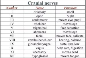
The twelve pairs of cranial nerves (I–XII):
They are: the olfactory nerve (I), the optic nerve (II), oculomotor nerve (III), trochlear nerve (IV), trigeminal nerve (V), abducens nerve (VI), facial nerve (VII), vestibulocochlear nerve (VIII), glossopharyngeal nerve (IX), vagus nerve (X), accessory nerve (XI), and hypoglossal nerve (XII).
Cranial nerves are generally named according to their structure or function.
Olfactory nerve (I) supplies smell
Facial nerve (VII) supplies motor innervation to the face.
The trochlear nerve (IV), named according to its structure, as it supplies a muscle that attaches to a pulley (Greek trochlea).
The trigeminal nerve (V) is named in accordance with its three components.
Th vagus nerve (X) is named for its wandering course (Latin: vagus).
Intracranial course of cranial nerves helps in the diagnosis of various intracranial lesions like brain tumors and intracranial arterial aneurysms.
The cell bodies of many of the neurons of most of the cranial nerves are contained in one or more nuclei in the brainstem.
Damage to these nuclei such as from a stroke or trauma can mimic damage to one or more branches of a cranial nerve.
The midbrain of the brainstem has the nuclei of the oculomotor nerve (III) and trochlear nerve (IV); the pons has the nuclei of the trigeminal nerve (V), abducens nerve (VI), facial nerve (VII) and vestibulocochlear nerve (VIII); and the medulla has the nuclei of the glossopharyngeal nerve (IX), vagus nerve (X), accessory nerve (XI) and hypoglossal nerve (XII).
The fibers of these cranial nerves exit the brainstem from these nuclei.
Some of the cranial nerves have sensory or parasympathetic ganglia which are located outside the brain.
The sensory ganglia are directly correspondent to dorsal root ganglia of spinal nerves and are known as cranial sensory ganglia.
Sensory ganglia exist for nerves with sensory function: V, VII, VIII, IX, X.
There are also parasympathetic ganglia, which are part of the autonomic nervous system for cranial nerves III, VII, IX and X.
The trigeminal ganglia of the trigeminal nerve (V) occupies a space in the dura mater called Trigeminal cave, and contains the cell bodies of the sensory fibers of the three branches of the trigeminal nerve.
The geniculate ganglion of the facial nerve (VII) is found as the nerve enters the facial canal.
The geniculate ganglion of the facial nerve (VII) contains the cell bodies of the sensory fibers of the facial nerve.
The superior and inferior ganglia of the glossopharyngeal nerve (IX), are located as the nerve passes through the jugular foramen and contain the cell bodies of the sensory fibers of this nerve.
The nodose ganglion which is the inferior ganglion of vagus nerve located below the jugular foramen and contains the cell bodies of the sensory fibers of the vagus nerve (X).
Exit sites of cranial nerves from the skull:
cribriform plate Olfactory nerve (I)
optic foramen Optic nerve (II)
superior orbital fissure Oculomotor (III)
Trochlear (IV)
Abducens (VI)
Trigeminal V1(ophthalmic)
foramen rotundum Trigeminal V2 (maxillary)
foramen ovale Trigeminal V3 (mandibular)
stylomastoid foramen Facial nerve (VII)
internal auditory canal Vestibulocochlear (VIII)
jugular foramen Glossopharyngeal (IX)
Vagus (X)
Accessory (XI)
hypoglossal canal Hypoglossal (XII)
After emerging from the brain, the cranial nerves travel within the skull, often passing through holes in the skull, called foramina, as they travel to their destinations.
Other nerves pass through bony canals.
Foramina and canals may contain more than one cranial nerve and may also contain blood vessels.
The olfactory nerve (I), is composed of many small separate nerve fibers, that passes through perforations in the cribriform plate part of the ethmoid bone.
The olfactory nerve fibers terminate in the upper part of the nasal cavity and function to convey impulses containing information about odors to the brain.
The optic nerve (II) passes through the optic foramen in the sphenoid bone as it travels to the eye, and conveys visual information to the brain.
The oculomotor nerve (III), trochlear nerve (IV), abducens nerve (VI) and the ophthalmic branch of the trigeminal nerve (V1) travel through the cavernous sinus into the superior orbital fissure, passing out of the skull into the orbit.
The III, IV, VI nerves control the small muscles that move the eye and also provide sensory innervation to the eye and orbit.
The trigeminal nerve (V2) maxillary division passes through foramen rotundum in the sphenoid bone to supply the skin of the middle of the face.
The mandibular division of the trigeminal nerve (V3) passes through foramen ovale of the sphenoid bone to supply the lower face with sensory innervation.
The mandibular division of the trigeminal nerve (V3) also sends branches to almost all of the muscles that control chewing.
The facial nerve (VII) and vestibulocochlear nerve (VIII) both enter the internal auditory canal in the temporal bone.
The facial nerve then reaches the side of the face by using the stylomastoid foramen, which is also in the temporal bone, and fibers then spread out to reach and control all of the muscles of facial expression.
The vestibulocochlear nerve reaches the control balance and hearing organs in the temporal bone.
The glossopharyngeal (IX), vagus (X) and accessory nerve (XI) all leave the skull via the jugular foramen to enter the neck.
The glossopharyngeal nerve provides innervation to the upper throat and the back of the tongue.
The vagus provides innervation to the muscles in the voicebox and continues downward to supply parasympathetic innervation to the chest and abdomen.
The accessory nerve controls the trapezius and sternocleidomastoid muscles in the neck and shoulder.
The hypoglossal nerve (XII) exits the skull using the hypoglossal canal in the occipital bone and reaches the tongue to control almost all of its muscles.
The cranial nerves provide motor and sensory innervation mainly to the structures within the head and neck.
The sensory innervation includes both general sensation such as temperature and touch, and special innervation such as taste, vision, smell, balance and hearing.
Olfactory nerve injury can cause an inability to smell, distortion in the sense of smell, or a distortion or lack of taste.
The optic nerve (II) transmits visual information to the brain.
Optic nerve damage leads to variable changes that depend on the location of the lesion.
Visual impairment may be homonymous hemianopsia or bitemporal hemianopsia.
Eye movement occurs by the III, IV, VI cranial nerves, and damage may affect the movement of the eyeball
The oculomotor nerve (III), trochlear nerve (IV) and abducens nerve (VI) coordinate eye movement.
With damage to these cranial nerves diplopia will likely occur because the movements of the eyes are no longer synchronized.
2242