 2213
2213
Numbered from 1 to 22, mainly based on size with 1 being the largest and 22 the smallest chromosome.
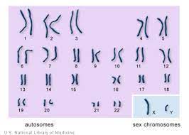
The first 22 pairs of chromosomes, called autosomes,are numbered from 1 to 22, from largest to smallest.
Human cells contain 23 pairs of chromosomes.
Chromosomes consist of the DNA sequence, and the proteins (such as histones) that are responsible for its packaging into chromosomes.
All cells, except for gametocytes, contain diploid genetic units.
The average chromosome contains 3-5000 genes and they range from a size of 1 kilobase to 2 megabases.
During meiosis recombination of genetic loci, or alleles, from one parent interchange with those from the other parent producing new combinations of alleles.
The likelihood that alleles will recombine during meiosis varies with the linear distance of one chromosome to another in the chromosomal sequence, this distance is referred to in centimorgans.
A centimorgan is the chromosomal distance over which there is a 1% chance that two alleles will undergo a crossover event during the meiosis process.
The recombination distance in the human genome is approximately 3000 centimorgans.
Crossover events during meiosis promotes genetic diversity among offspring and indicates that specific alleles are inherited together.
Threadlike structures of nucleic acids and protein found in the nucleus of most living cells, carrying genetic information in the form of genes.
Sometimes referred to as the individualized portions of chromatin in cells, either visible or not under light microscopy.
Sometimes referred to as the individualized portions of chromatin during cell division, visible under light microscopy due to high condensation.
A chromosome is a DNA molecule with part or all of the genetic material of an organism.
Most eukaryotic chromosomes include packaging proteins which, bind to and condense the DNA molecule to prevent it from becoming an unmanageable tangle.
Normally visible under a light microscope only when the cell is undergoing the metaphase of cell division, where all chromosomes are aligned in the center of the cell in their condensed form.
Every chromosome is copied once in S phase, and the copy is joined to the original by a centromere, resulting either in an X-shaped structure if the centromere is located in the middle of the chromosome or a two-arm structure if the centromere is located near one of the ends.
The original chromosome and the copy are called sister chromatids.
The X-shape structure formed in metaphase is called a metaphase chromosome, and it is a highly condensed.
Chromosomal recombination during meiosis plays a significant role in genetic diversity.
Through chromosomal instability and translocation, the cell may undergo mitotic catastrophe and die.
Chromosomal mutations in the cell can allow it to inappropriately evade apoptosis and lead to the progression of cancer.
Chromosomes are composed of chromatin fiber, which are made of nucleosomes, which are packaged by proteins into a condensed structure called chromatin.
Chromatin contains the vast majority of DNA and a small amount can be found in the mitochondria.
Chromatin is present in most cells.
Chromatin allows the long DNA molecules to fit into the cell nucleus.
During cell division chromatin increasingly condensed to form chromosomes.
During cellular division chromosomes are replicated, divided, and passed successfully to their daughter cells so as to ensure the genetic diversity and survival of their progeny.
Unduplicated chromosomes are single double helixes.
Duplicated chromosomes contain two identical copies, called chromatids or sister chromatids, joined by a centromere.
Chromosomes may exist as either duplicated or unduplicated.
Each chromosome has one centromere, with one or two arms projecting from the centromere, although, under most circumstances, these arms are not visible as such.
During interphase, when the cell is not dividing, there are two types of chromatin that can be distinguished:
Euchromatin, which consists of DNA that is actively being expressed as protein.
Heterochromatin, which consists of mostly inactive DNA.
Heterochromatin can be further distinguished into two types:
Constitutive heterochromatin
Facultative heterochromatin
Each chromosome is made up of two chromatids, chromosomal arms, which are joined to each other at a small constricted region called the centromere, the primary constriction.
Chromatids are conjoined twins the result of DNA replication.
The centromere helps the chromatids attach to the spindle fibers during cell division.
The centromere is also concerned with the anaphase movement of the chromosomes, by which the spindle fibers pull the chromatids to the two opposite poles by their contraction during anaphase.
In certain chromosomes there is a secondary constriction as well.
The two chromatids are made up of very thin chromatin fibers which are made up of 40% DNA and 60% histone proteins
Each chromatin fiber consists of one DNA helix coiled around eight histone molecules like a loop, and is referred to as a nucleosome.
Nucleosomes pack tighter, during condensation required to get to metaphase.
Within the primary constriction there is a clear zone called Centromere.
The centromere with the DNA and histone proteins bound to them form a disc shaped structure called kinetochore.
In the early stages of mitosis or cell division, (meiosis) , the chromatin double helix become more and more condensed, stoping transcription and becoming a compact transportable form.
This compact form is the classic four arm structure, a pair of sister chromatids attached to each other at the centromere.
The shorter arms are called p arms and the longer arms are called q arms.
Chromosomes are further sub-divided into many bands that are numbered.
For example, “chromosome 3p21.3” refers to band 21 on the short arm of chromosome 3.
The numbered bands specify the location of the thousands of genes that are present on each chromosome.
The compact form is when individual chromosomes are visible with an optical microscope.
During mitosis, microtubules grow from centrosomes located at opposite ends of the cell and also attach to the centromere at specialized structures called kinetochores, one of which is present on each sister chromatid.
Microtubules pull the chromatids apart toward the centrosomes, so that each daughter cell inherits one set of chromatids.
Once the cells have divided, the chromatids are uncoiled and DNA can again be transcribed.
Chromosomes are structurally highly condensed, enabling these giant DNA structures to be contained within a cell nucleus.
Chromosomes are divided into two types:
autosomes-body chromosomes
allosomes -sex chromosomes
Genetic traits are linked to a person’s sex and are passed on through the sex chromosomes.
The autosomes contain the rest of the genetic hereditary information.
All cells have 23 pairs of chromosomes (22 pairs of autosomes and one pair of sex chromosomes), giving a total of 46 per cell.
Cells also have many hundreds of copies of the mitochondrial genome.
Estimated number of genes on each human chromosome:
1 2000
2 1300
3 1000
4 1000
5 900
6 1000
7 900
8 700
9 800
10 700
11 1300
12 1100
13 300
14 800
15 600
16 800
17 1200
18 200
19 1500
20 500
21 200
22 500
X (sex chromosome) 800
Y (sex chromosome) 50
Total 21,000 genes
Most eukaryotes are diploid, with 22 different types of autosomes, each present as two homologous pairs, and two sex chromosomes.
This gives 46 chromosomes in total.
Gametes, reproductive cells, are haploid, having one set of chromosomes.
Gametes are produced by meiosis of a diploid germ line cell.
During meiosis, the matching chromosomes of father’s and mother’s small parts can crossover, and thus create new chromosomes that are not inherited solely from either parent.
When fertilization occurs and a male and a female gamete merge, a new diploid organism is formed.
The karyotype is the characteristic chromosome complement and are often highly variable.
Karyotype variations include:
1. variation between the two sexes
2. variation between the germ-line and soma
3. variation between members of a population, due to balanced genetic polymorphism
4. geographical variation between races
5. mosaics or otherwise abnormal individuals.
Variation in karyotype may occur during development from the fertilized egg.
The technique of determining the karyotype is usually called karyotyping.
In karyotyping cells can be locked part-way through division, in metaphase, in vitro with colchicine.
These cells are then stained, photographed, and arranged into a karyogram, with the set of chromosomes arranged, autosomes in order of length, and sex chromosomes at the end.
Humans have special sex chromosomes, in contrast to autosomes, and these are XX in females and XY in males.
Chimpanzees, have 48 chromosomes as do the other great apes: in humans two chromosomes fused to form chromosome 2.
Chromosomal aberrations are caused by the disruptions in the normal chromosomal content of a cell and are a major cause of genetic conditions in humans.
Most chromosomal aberrations have little to no effect.
Some chromosome abnormalities do not cause disease in carriers, such as translocations, or chromosomal inversions.
Abnormal numbers of chromosomes or chromosome sets, called aneuploidy, may be lethal or may give rise to genetic disorders.
The gain or loss of DNA from chromosomes can lead to a genetic disorders.
In Down syndrome, there are three copies of chromosome 21.
Cri du chat, which is caused by the deletion of part of the short arm of chromosome 5.
Edwards syndrome, or trisomy-18, the second most common trisomy.
Klinefelter syndrome (XXY).
Patau Syndrome, also called D-Syndrome or trisomy-13.
Pallister–Killian syndrome.
Triple-X syndrome (XXX).
Turner syndrome (X instead of XX or XY).
In Turner syndrome, female sexual characteristics are present but underdeveloped.
XYY syndrome.
XXX syndrome
Aneuploidy is associated with lifestyle, environmental and/or occupational hazards, tobacco smoking, exposure to benzene, insecticides, perfluorinated compounds and increased damage to sperm.
Chromosome 6 contains the major histocompatibility complex, which contains over 100 genes related to the immune response, and plays a vital role in organ transplantation.