 Butterfly vertebra is also known as sagittal cleft vertebra, is a rare congenital spinal anomaly characterized by the presence of a sagittal cleft within a vertebral body, giving it a butterfly-like appearance on imaging.
This condition arises due to incomplete fusion of the lateral halves of a vertebra during embryonic development.
It is often asymptomatic, butterfly vertebrae may occasionally be associated with spinal deformities or syndromic conditions.
The vertebral column develops from paired somites during embryogenesis: the right and left halves of each vertebra fuse in the midline to form a complete vertebral body.
In butterfly vertebrae, this fusion process is disrupted, leading to a persistent sagittal cleft.
The defect is usually filled with fibrous or cartilaginous tissue, and the two halves of the vertebral body may remain connected by this intervening soft tissue.
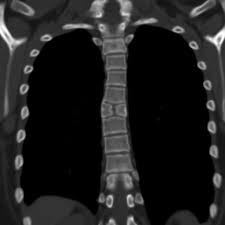
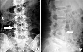
The condition is most commonly observed in the thoracic and lumbar spine.
It can occur at any spinal level.
The degree of clefting varies, with a spectrum of appearances on imaging studies.
The vertebra appears to be divided into two symmetrical halves, separated by a vertical lucency that represents the cleft.
The lateral portions of the vertebral body often appear sclerotic, and the shape resembles a butterfly when viewed in the anteroposterior projection.
CT imaging provides greater detail regarding the bony anatomy, including the extent of clefting and the composition of the intervening tissue.
MRI: Useful for evaluating associated spinal cord abnormalities or adjacent soft tissue changes.
The sagittal cleft may appear as a hyperintense signal on T2-weighted MRI images, representing cartilaginous or fibrous material.