 The B-cell receptor (BCR) is a transmembrane protein on the surface of a B cell.
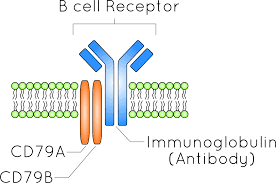
A B-cell receptor includes both CD79 and the immunoglobulin.
The B- cell receptor extends both outside the cell (above the plasma membrane) and inside the cell (below the membrane).
A B-cell receptor is composed of a membrane-bound immunoglobulin molecule and a signal transduction moiety.
The former forms a type 1 transmembrane receptor protein, and is typically located on the outer surface of these lymphocyte cells.
Through biochemical signaling and by physically acquiring antigens from the immune synapses, the BCR controls the activation of the B cell.
B cells are able to engage biochemical modules for receptor clustering, cell spreading, generation of pulling forces, and receptor transport, which eventually culminates in endocytosis and antigen presentation.
B cells’ mechanical activity adheres to a pattern of negative and positive feedbacks that regulate the quantity of removed antigen by manipulating the dynamic of BCR–antigen bonds directly.
By grouping and spreading increase the relation of antigen with BCR, thereby proving sensitivity and amplification.
Pulling forces can delink the antigen from the BCR, thus testing the quality of antigen binding.
The BCR for an antigen is a significant sensor that is required for B cell activation, survival, and development.
A B cell is activated by its first encounter with an antigen that binds to its receptor, resulting in cell proliferation and differentiation to generate a population of antibody-secreting plasma B cells and memory B cells.
The B cell receptor (BCR) has two crucial functions upon interaction with the antigen.
One function is signal transduction, involving changes in receptor oligomerization.
The second function is to mediate internalization for subsequent processing of the antigen and presentation of peptides to helper T cells.
The first checkpoint in the development of a B cell is the production of a functional pre-BCR, which is composed of two surrogate light chains and two immunoglobulin heavy chains, which are normally linked to Ig-α (or CD79A) and Ig-β (or CD79B) signaling molecules.
Each B cell, produced in the bone marrow, is highly specific to an antigen.
The general structure of the B cell receptor includes a membrane-bound immunoglobulin molecule and a signal transduction region.
Disulfide bridges connect the immunoglobulin isotype and the signal transduction region.
The B-cell receptor is composed of two parts:
A membrane-bound immunoglobulin molecule of one isotype (IgD, IgM, IgA, IgG, or IgE).
Signal transduction moiety: a heterodimer called Ig-α/Ig-β (CD79), bound together by disulfide bridges.
Each member of the dimer spans the plasma membrane and has a cytoplasmic tail bearing an immunoreceptor tyrosine-based activation motif,
BCR complex consists of an antigen-binding subunit known as the membrane immunoglobulin (mIg), which is composed of two immunoglobulin light chains (IgLs) and two immunoglobulin heavy chains (IgHs) as well as two heterodimer subunits of Ig-α and Ig-β.
Within the BCR, the part that recognizes antigens is composed of three distinct genetic regions, referred to as V, D, and J.
There are a number of genes that encode each of these regions in the genome and can be joined in various ways to generate a wide range of receptor molecules:
Through this process,the body finds a way of producing multiple different combinations of antigen-recognizing receptor molecules.
Heavy chain rearrangement of the BCR entails the initial steps in the development of B cell.
BCRs have distinctive binding sites that rely on the complementarity of the surface of the epitope and the surface of the receptor.
Mature B cells can only survive in the peripheral circulation for a limited time when there is no specific antigen.
This is because when cells do not meet any antigen within this time, they will go through apoptosis.
In the peripheral circulation, apoptosis is important in maintaining an optimal circulation of B-lymphocytes.
The BCR for antigens are almost identical to secreted antibodies.
Signaling pathways of the B cell receptor
The physiology of B cells is intimately connected with the function of their B-cell receptor.
The BCR signaling pathway is initiated when the mIg subunits of the BCR bind a specific antigen.
The B-cell receptor has been shown to be involved in the pathogenesis of various B cell derived lymphoid cancers.
Stimulation by antigen binding may contribute to the proliferation of malignant B cells, but increasing evidence implicates antigen-independent self-association of BCRs as a key feature in a growing number of B cell neoplasias.
B cell receptor signalling is currently a therapeutic target in various lymphoid neoplasms.
It has been shown that BCR signaling is synchronised with CD40 pathway activation provided by B-T cell interactions, and this seems to be essential to trigger proliferation of leukemic B cells.
Plays an important part in the development and maturation of B-cells.
BCR signaling please roll in B-cell activation, proliferation, and survival.
BCR activation provides one of the most important survival signals to CLL B-cells.
B-cell receptors assist with binding, internalization, and processing of an antigen.
Such signals are transduced through a variety of kinases including: LYN, PI3K, SYK, and BTK, and results in the phosphorylation of PLC-gamma2 and induction of downstream second messengers modulating cell survival regulators.
BCR signaling in CLL cells is activated in secondary lymphatic organs by soluble or cell-surface antigens, which can be autoantigens.
Homotypic interactions between BCR molecules result in autonomous BCR signaling.
BCR activation causes downstream signal- ing through spleen tyrosine kinase (SYK), Bruton’s tyrosine kinase (BTK), and phosphatidylinositol 3-kinase (PI3K), which are the targets of the BTK inhibitors (BTKi) ibrutinib and acalabrutinib and the PI3K inhibitors (PI3Ki) idelalis- ib and duvelisib. SYK, BTK, and PI3K also transmit signaling from chemokine receptors, such as CXCR4 and CXCR5, which regulate tissue homing and retention of CLL cells.
Stimulation of B-cell receptors induces the activation of multiple enzymes, including Bruton tyrosine kinase (BTK) which is part of the B-cell receptor signaling pathway that communicates with other cells of the immune system and results in B-cell proliferation and activation.
Abnormal BCR signaling is implicated as a key pathway in leukemogenesis.
Kinase inhibitors such as Ibrutinib and Idelalisib target different mediated of BCR signaling.
By targeting the different kinases involved in the BCR pathway survival signals can be prevented resulting in apoptosis of cancer cells.
The disruption of the signals can result in mobilization of malignant B-cells in lymph nodes and bone marrow, allowing access to the immune system.