
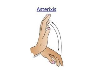
 Asterixis, often called a “flapping tremor” or “liver flap,” is an involuntary movement disorder.
Asterixis Is caused by a brief, sudden loss of muscle tone when a person tries to hold a position.
Asterixis is a disorder of motor control defined as sudden, brief, arrhythmic lapses of sustained posture caused by involuntary interruption of muscle contraction.
It is a specific form of negative myoclonus — a momentary loss of muscle tone in agonist muscles followed by a compensatory jerk of antagonist muscles.
This makes the hands flap or jerk downward and then spring back up.
Main CausesAsterixis is usually a sign of a metabolic problem that affects how the brain controls muscle posture.
Etiologies Metabolic/toxic (most common): Hepatic failure Renal failure / uremia Respiratory failure (hypercapnia or hypoxia) Electrolyte disturbances: hypokalemia, hypo/hypermagnesemia, hypercalcemia, hypophosphatemia, hyponatremia, hypoglycemia Hyperammonemia (with or without liver dysfunction) Sepsis-associated encephalopathy, hyperviscosity, polycythemia Drug-induced (phenytoin is the most frequently reported): Antiseizure meds: valproate, carbamazepine, phenytoin, gabapentin, lamotrigine, phenobarbital, pregabalin, primidone Antipsychotics: clozapine, olanzepine, risperidone Others: lithium, benzodiazepines, metoclopramide, ceftazidime, amantadine, opioids, amphotericin B Valproate, phenytoin, and carbamazepine can worsen asterixis by raising serum ammonia Structural brain lesions: Stroke (ischemic, hemorrhagic, TIA) — prevalence ~1.9% in post-stroke patients Subdural/epidural hematoma, tumors, abscess Lesion locations: thalamus (most common), midbrain, pons, cerebellum, frontoparietal cortex, basal ganglia, internal capsule
Other: Wilson’s disease, Creutzfeldt-Jakob disease, cerebral malaria, trypanosomiasis, episodic ataxia type 1 (KCNA1 mutation), post-thalamotomy
Asterixis may be unilateral, bilateral, or asynchronous, with irregular frequency and amplitude.
Typically asymptomatic ad not spontaneously reported by patients, but is detected on exam.
Can affect wrists, fingers, ankles, proximal limbs, neck, face, tongue, lips, eyelids, and trunk
Bilateral asterixis → more commonly metabolic/drug-related
Unilateral asterixis → suggests a contralateral structural brain lesion (thalamus is the most common site, ~54% of cases)
Clinical frequency ~0.5–2 Hz; EMG shows pauses of 35–200 ms in affected muscles
Examination
Holding arms outstretched, spread fingers, dorsiflex wrists, close eyes, and open mouth.
Observe for brief downward flaps with quick return to posture.
Wait at least 30 seconds before concluding it is absent as there is a significant latent period.
If not apparent, softly sweep-extend the wrist.
Lower limb: Patient supine with knees bent, feet flat, let legs fall to sides — observe for flapping at hips with knees springing back together.
Alternative methods: Squeeze examiner’s hand, or squeeze a partially inflated BP cuff as readings jump wildly in asterixis.
West Haven Grading for Hepatic Encephalopathy
Asterixis appears at Grade 2 (with disorientation, lethargy, paratonia, dysarthria) and disappears at Grade 4 (coma).
It carries prognostic value — ~56% of patients with alcohol-associated liver disease and asterixis had poor outcomes.
Its precise mechanism remains unknown.
Proposed mechanisms include: Disturbance of ascending activating systems in encephalopathy Dysfunction of thalamo-motor-cortical coupling and sensorimotor integration (parietal/midbrain regions) Abnormal cortical motor activity with negative sharp waves in the contralateral central area The dentato-rubro-thalamic circuitry is the principal pathway implicated Postural control involves vestibulospinal, reticulospinal, and rubrospinal tracts modulated by supratentorial structures, with convergence at the ventrolateral thalamus
Differential Diagnosis Tremor — rhythmic oscillation vs. asterixis’s arrhythmic antigravitational lapses Clonus — rhythmic, stretch-reflex induced, associated with hyperreflexia Dyskinesia — affects throughout movement vs. asterixis being posture-related Pseudoasterixis — brief voluntary action tremors; patients are aware of the twitching Epileptic negative myoclonus — EMG silent period <500 ms without antagonist contraction; may require EEG to differentiate; ethosuximide or levetiracetam may be effective
Evaluation:
CBC, electrolytes, glucose, renal/liver function panels, arterial blood gas, drug levels/toxicology, ammonia. Neuroimaging (MRI/MRA) for unilateral asterixis. EEG may show generalized slowing (uremic), triphasic waves (hepatic), or epileptiform discharges (epileptic negative myoclonus). EMG if phenomenological differentiation is difficult.
Treatment Asterixis is usually reversible with treatment of the underlying cause : Hepatic encephalopathy: lactulose (titrate to 2–4 loose stools/day), rifaximin (400 mg PO TID), L-ornithine L-aspartate, dietary modification with vegetable protein preferred Uremic: dialysis, renal transplantation, dialysis dose adjustment Drug-induced: identify and discontinue/adjust the offending agent; L-carnitine for valproate-related hyperammonemia Wilson’s disease: low-copper diet, D-penicillamine, trientine, zinc; liver transplantation as rescue therapy Structural lesions: treat the underlying lesion (e.g., stroke, hematoma, tumor) Deep brain stimulation: GPi-DBS improved asterixis in a patient with a KMT2B mutation, suggesting shared dystonia/asterixis mechanisms