
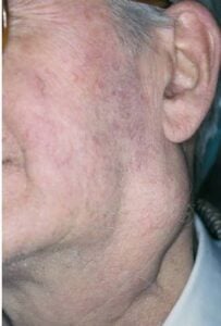
 A Warthin’s tumor is a common, non-cancerous growth that develops in the salivary glands, almost always in the parotid gland near the ear.
It is also called papillary cystadenoma lymphomatosum or adenolymphoma, and is a benign salivary gland neoplasm arising almost exclusively in the parotid gland, where it is the second most common tumor after pleomorphic adenoma, comprising roughly 5–15% of parotid tumors.
It is defined histologically by a bilayered oncocytic lesion that is mitochondria-rich epithelium forming papillary-cystic structures supported by a dense lymphoid stroma with germinal centers.
Malignant transformation is extremely rare (<0.1–0.3%).
It is strongly linked to smoking, usually affects older adults, and often presents as a slow-growing, painless lump.
Typically presents in the 6th–7th decades with a male predominance, though the gender gap is narrowing with rising smoking rates in women.
Smoking is the dominant risk factor, with reported odds 7- to 40-fold higher in smokers versus nonsmokers.
Additional associations include aging, radiation exposure, chronic inflammation, and—more recently reported—obesity/metabolic syndrome.
Presents a slow growth, usually near the angle of the jaw or lower ear.
Usually a painless, slowly growing, soft-to-firm mass in the tail of the parotid lower pole.
Less commonly causes ear pain, tinnitus, or facial nerve branch irritation.
Average growth is slow (~8%/year; mean doubling time ~9 years), though occasional rapid enlargement occurs.
Most people feel no pain at all.
Smokers have a much higher chance of getting this tumor.
Multiple or both sides: About 10% to 20% of cases can happen on both sides or as multiple lumps in ~12–20% of cases, higher than other benign parotid tumors.
Diagnosis
Ultrasound or CT scan help show where the lump is and if it has fluid inside.
Ultrasound plus fine-needle aspiration cytology (FNAC) is the mainstay.
The classic cytologic triad is oncocytes, a polymorphous lymphocyte population, and a “grungy” granular/proteinaceous background; when all three are present, specificity is high.
FNAC accuracy is strong in experienced hands with a reported sensitivity ~78–98%, specificity ~96–98%, high PPV, though large cystic components can yield nondiagnostic, paucicellular samples.
Imaging pitfall: WT is frequently FDG-avid on PET/CT and may show restricted diffusion on MRI, mimicking malignancy or nodal metastasis—important in head and neck cancer staging.
On multiphase CT, WT characteristically shows early peak enhancement with delayed washout.
Fine needle test confirms it is not cancer.
Treatment Options
Surgery: Doctors often remove the lump or a small part of the gland.
Surgery is indicated for symptomatic, enlarging, diagnostically uncertain, or patient-preference cases.
Trends favor tissue-preserving approaches (extracapsular dissection, partial superficial parotidectomy, or enucleation) over traditional superficial/total parotidectomy, given WT’s well-demarcated intact capsule.
Surgical complications include great auricular nerve sensory deficit (common), permanent facial nerve paralysis (~2–8%), Frey syndrome, salivary fistula, and hematoma.
Watch and wait.
Active surveillance is a safe, increasingly favored option for small, asymptomatic, cytologically confirmed WT—particularly in elderly or comorbid patients—given the very low malignant potential and slow growth.
In observational cohorts, growth is minimal and progression to malignancy was not observed.
The chance of the tumor coming back after surgery is very low.
Smoking cessation is relevant, as continued smoking is associated with metachronous contralateral tumors.