The vestibular system is a sensory system that creates the sense of balance and spatial orientation for the purpose of coordinating movement with balance.
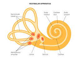
Together with the cochlea, a part of the auditory system, it constitutes the labyrinth of the inner ear.
Movements consist of rotations and translations.
The vestibular system comprises two components: the semicircular canals, which indicate rotational movements; and the otoliths, which indicate linear accelerations.
The vestibular system sends signals primarily to the neural structures that control eye movement; these provide the anatomical basis of the vestibulo-ocular reflex (standardofcare.com/vestibulo-ocular-reflex) which is required for clear vision.
Signals are also sent to the muscles that keep one upright and in general control posture.
The vestibular system sends signals that provide the anatomical means required to enable one to maintain its desired position in space.
The brain uses information from the vestibular system in the head and from proprioception throughout the body to understand its body’s dynamics and kinematics.
That includes its position and acceleration from moment to moment.
The semicircular canal system detects rotational movements.
The vestibular system contains three semicircular canals in each labyrinth.
They are approximately orthogonal, at right angles, to each other, and are the horizontal,or lateral, the anterior semicircular canal, or superior, and the posterior, or inferior semicircular canal.
The anterior and posterior canals may collectively be called vertical semicircular canals.
Movement of fluid within the horizontal semicircular canal corresponds to rotation of the head around a vertical axis, as in a pirouette.
The anterior and posterior semicircular canals detect rotations of the head in the sagittal plane, as when nodding, and in the frontal plane, as when cartwheeling.
Both anterior and posterior canals are oriented at approximately 45° between frontal and sagittal planes.
The movement of semicircular fluid pushes on a the cupula which contains hair cells that transduce the mechanical movement to electrical signals.
Push-pull systems
The semicircular canals are arranged in such a way that each canal on the left side has an almost parallel counterpart on the right side.
Each of these three pairs works in a push-pull fashion: when one canal is stimulated, its corresponding partner on the other side is inhibited, and vice versa.
As a result of this push-pull system it is possible to sense all directions of rotation.
The right horizontal canal gets stimulated during head rotations to the right.
The left horizontal canal gets stimulated by head rotations to the left.
The vestibulo-ocular reflex occurs when a rotation of the head is detected, triggering an inhibitory signal to the extraocular muscles on one side and an excitatory signal to the muscles on the other side, resulting is a compensatory movement of the eyes.
The vestibular-ocular reflex (VOR) is a reflex eye movement that stabilizes images on the retina during head movement.
VOR produces an eye movement in the direction opposite to head movement, thus preserving the image on the center of the visual field: when the head moves to the right, the eyes move to the left, and vice versa.
There is a slight head movement present all the time, and the VOR is important to stabilizing vision: patients whose VOR is impaired find it difficult to read because they cannot stabilize the eyes during small head tremors.
The VOR reflex does not depend on visual input and works even in total darkness or when the eyes are closed.
This VOR reflex, combined with the push-pull principle described above, forms the physiological basis of the Rapid head impulse test or Halmagyi-Curthoys-test.
The rapid head impulse: the head is rapidly and forcefully moved to the side while observing whether the eyes keep looking in the same direction.
The velocity of the eyes must be opposite to the velocity of the head to maintain clear vision.
Signals from the vestibular system also project to the cerebellum to keep the VOR effective, and to different areas in the cortex.
The vestibular nuclei on either side of the brainstem exchange signals regarding movement and body position.
The vestibular nuclei of the brainstem send signals to the cerebellum.
The signals sent to the cerebellum are relayed back as muscle movements of the head, eyes, and posture.
The vestibular nuclei of the brainstem send signals to the nuclei of cranial nerves III, IV, and VI causing the vestibular-ocular reflex.
The vestibular-ocular reflex allows the eyes to fix on a moving object while staying in focus.
The vestibular nuclei of the brainstem send signals to the
reticular formation, indicating the new posture the body has taken on, and how to adjust circulation and breathing due to body position.
The vestibular nuclei of the brainstem send signals to the spinal cord, allowing quick reflex reactions to both the limbs and trunk to regain balance.
The vestibular nuclei of the brainstem send signals to the thalamus, allowing for head and body motor control as well as being conscious of body position.
The semicircular canals respond to rotations.
The otolithic organs sense linear accelerations.
There are two otolithic organs on each side, one called the utricle, the other called the saccule.
The utricle and saccule contain a patch of hair cells and supporting cells called a macula.
Each hair cell of a macula has forty to seventy stereocilia and one true cilium called a kinocilium.
The tips of cilia are embedded in an otolithic membrane.
The otolithic membrane is weighted down with protein-calcium carbonate granules called otoconia.
Otoconia add to the weight and inertia of the membrane and enhance the sense of gravity and motion.
With the head erect, the otolithic membrane bears directly down on the hair cells and stimulation is minimal.
When the head is tilted, the otolithic membrane sags and bends the stereocilia, stimulating the hair cells.
Orientation of the head causes a combination of stimulation to the utricles and saccules.
The brain interprets head orientation by comparing these inputs to each other and other input from the eyes and stretch receptors in the neck, allowing detection of whether the head is tilted or the entire body is tipping.
Otolithic organs sense how quickly you are accelerating forward or backward, left or right, or up or down.
Most of the utricular signals elicit eye movements.
The majority of the saccular signals projects to muscles that control our posture.
The semicircular canals interpret rotation signals.
Otolith signals are caused by linear movements and also gravity.
Humans can sense head tilting and linear acceleration even in dark environments because of the orientation of two groups of hair cell bundles on either side of the striola.
The hair cells on opposite sides move with mirror symmetry, when one side is moved, the other is inhibited.
The opposing effects caused by a tilt of the head cause varying sensory inputs from the hair cell bundles allowing humans to tell which way the head is tilting.
This sensory input is sent to the brain, which responds with corrective actions to the nervous and muscular systems ensuring balance and awareness are maintained.
When the vestibular system is stimulated without any other inputs, one experiences a sense of self-motion.
Compared to the other senses of vision, touch and audition, vestibular input is perceived with delay.
Diseases of the vestibular system usually induce vertigo and instability or loss of balance, often accompanied by nausea.
The most common vestibular diseases in humans are vestibular neuritis, a related condition called labyrinthitis, Ménière’s disease, and BPPV.
The vestibular system’s function can be affected by tumors on the vestibulocochlear nerve, an infarct in the brain stem or in cortical regions related to the processing of vestibular signals, and cerebellar atrophy.
When the vestibular system and the visual system report incongruous results, nausea often occurs.
When the vestibular system reports movement but the visual system reports no movement, the motion disorientation is often called motion sickness/seasickness/car sickness.
When a person is in a zero-gravity environment or during a virtual reality session, the disoriented sensation is often called space sickness or space adaptation syndrome.
Alcohol can also cause alterations in the vestibular system for short periods, resulting in vertigo and possibly nystagmus due to the altered viscosity of the blood and the endolymph during the consumption of alcohol: positional alcohol nystagmus.
Positional alcohol nystagmus1(PAN1) The alcohol concentration is higher in the blood than in the vestibular system, hence the endolymph is relatively dense.
Positional alcohol nystagmus II PAN II – The alcohol concentration is lower in the blood than in the vestibular system, hence the endolymph is relatively dilute.
PAN I will result in subjective vertigo in one direction and typically occurs shortly after ingestion of alcohol when blood alcohol levels are highest.
PAN II will eventually cause subjective vertigo in the opposite direction.
This occurs several hours after ingestion and after a relative reduction in blood alcohol levels.
Benign paroxysmal positional vertigo (BPPV) resulting in acute symptoms of vertigo and is caused when pieces that have broken off otoliths have slipped into one of the semicircular canals.
In most cases, it is the posterior canal that is affected.
These particles can shift and create a fluid wave which displaces the cupula of the canal affected, which leads to dizziness, vertigo and nystagmus.
Vestibular dysfunction has also been found to correlate with cognitive and emotional disorders, including depersonalization and derealization.