
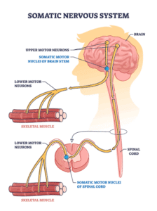
 Upper motor neurons (UMNs) start in the motor cortex and travel down the brainstem and spinal cord (corticospinal and corticobulbar tracts).
They don’t leave the CNS.
They synapse on lower motor neurons, mostly through interneurons.
Lower motor neurons (LMNs) have cell bodies in the brainstem or spinal cord anterior horn.
Their axons leave the CNS through cranial or spinal nerves and directly innervate skeletal muscle.
They are the final common pathway.
Upper motor neuron (UMN) and lower motor neuron (LMN) lesions produce distinct, largely opposite clinical signatures, and distinguishing between them is the first step in localizing any weakness.
UMNs originate from Betz cells in layer V of the primary motor cortex and descend via the corticospinal/corticobulbar tracts.
LMNs are the brainstem cranial motor nerve nuclei and anterior horn cells that directly innervate skeletal muscle.
UMN lesions release the LMN from cortical inhibition, producing a spastic/hyperreflexic picture, whereas LMN lesions denervate muscle, producing a flaccid/atrophic picture.
UMN: stroke, spinal cord injury above the lesion level, multiple sclerosis
LMN: peripheral neuropathy, polio, Guillain-Barré syndrome, nerve root compression
Both: ALS, which is why it shows a mix of spasticity and atrophy with fasciculations
Note that acute UMN injuries, like a fresh stroke or spinal cord injury, can initially look flaccid (spinal shock) before spasticity develops.
Characteristic UMN signs: slowing of fine, skilled movements like rapid finger or foot tapping, increased tone, spasticity, hyperreflexia, and pathologic reflexes including Babinski, Hoffmann’s sign, jaw jerk, snout reflex, and pathologic spread of reflexes.
Preserved (or brisk) reflexes in a limb that is weak and atrophic is itself a clue to concurrent UMN involvement.
Characteristic LMN signs: weakness of the involved muscle, muscle atrophy, fasciculations, decreased tone/flaccidity, and hyporeflexia.
The UMN-vs-LMN distinction anchors lesion localization — pure LMN findings point toward the anterior horn cell, root, plexus, peripheral nerve, neuromuscular junction, or muscle, and make brain/cord localization improbable.
Motor neuron diseases are classically framed along this spectrum: combined UMN + LMN = amyotrophic lateral sclerosis (ALS); pure UMN = primary lateral sclerosis and hereditary spastic paraplegia; pure LMN = progressive muscular atrophy, spinal muscular atrophies, and multifocal motor neuropathy.