Type 1 diabetes, also known as juvenile diabetes.
More than 8.4 million people worldwide live with type 1 diabetes.
Approximately 0.5 million new cases of type one diabetes are diagnosed per year worldwide.
Type 1 diabetes is most commonly diagnosed between ages 10 and 14 years.
In 2021 the median age of diagnosis was 29 years worldwide and the median age at diagnosis in the US is about 24 years.
However, more than half the cases of type 1 diabetes are diagnosed in adulthood; 62% of recent new cases were in patients over the age of 20 years.
There are genetic, immune, and metabolic differences between childhood and adult onset type 1 diabetes.
The mean age of a person living with type 1 diabetes is now 40 years-due to later age at diagnosis, and longer lifespan.
Onset of type 1 diabetes is more common in fall in winter, likely due to viral infections, which precipitates hyperglycemia.
It is a form of diabetes in which very little or no insulin is produced by the pancreas.
It is caused by the progressive autoimmune destruction of insulin producing pancreatic beta cells initiated, as yet by unconfirmed triggering events, such as a viral infection, in persons who are genetically predisposed.
Autoimmune mediated dysfunction of insulin producing beta cells occurs in pancreatic islets and lead to dysglycemia and lifelong dependence on insulin therapy.
The auto immune process in type 1 diabetes involves adaptive and innate immune responses.
Type 1 diabetes can be diagnosed in presymptomatic stages via detection of two or more islet autoantibodies.
Pharmacologic intervention can delay the onset of clinical, symptomatic type 1 diabetes, thereby altering the underlying disease course:suggesting increase utilization of screening even beyond genetically selected populations.
Stage I is defined by the presence of two or more islet autoantibodies with normoglycemia and stage 2 by the presence of two or more islet autoantibodies and abnormal glucose metabolism.
Stage 3 marks the clinical diagnosis of type 1 diabetes, characterized by hyperglycemia.
Islet specific auto antibodies direct CD8 T cells to the pancreatic islet where they bind to the antigen specific class 1 major histocompatibility complex on the beta cell surface, causing release of performin and granzymeB, which leads to beta cell apoptosis.
People with type 1 diabetes may have defects in the beta cell, such as an altered prohormone processing, that facilitate development of auto immunity.
In type 1 diabetes, the endoplasmic reticulum within the beta cell is responsible for insulin synthesis and protein folding, accumulates misfolded proteins, including abnormal accumulation of proinsulin, which contributes to beta cell death, and the release of islet auto antigens into the peripheral circulation.
Type one diabetes can be detected prior to symptom onset by the presence of multiple islet autoantibodies.
Continued beta cell destruction leads to hyperglycemia, which may take many years.
Patients with type one diabetes, have a life expectancy that is approximately 13 years shorter than that of the general population.
The primary cause of a shortened life expectancy is cardiovascular disease.
There is a close association between levels of glycated hemoglobin, and the risk of death from cardiovascular disease.
Nearly one in three persons with type one diabetes will go on to have diabetic kidney disease, an independent risk factor for cardiovascular events and premature death.
Studies show that in patients with a glycated hemoglobin level of 6.9% or lower, had a risk of death from cardiovascular causes that was greater by a factor of two than that of non-diabetic controls.
To prevent long-term microvascular and macrovascular complications of type one diabetes the required insulin dose is adjusted to reach glucose levels of hemoglobin A1c less than 7% and a percentage of time spent in the target glucose range of 70 to 180 mg/dL of more than 70%.
Approximately 75% persons with type one diabetes, do not have a glycated hemoglobin level of less than 7% and in patients with the hemoglobin level: if glycated hemoglobin level is 7% or higher there is an increased risk of retinopathy, neuropathy, nephropathy, cardiovascular disease, and early death.
A systemic review and meta-analysis of more than 214,000 patients with type one diabetes showed a relative risk of cardiovascular events that was twice as high for women as it was for men.
Autoreactive, CD8, positive T cells in type one diabetes bind to, and activated by autoantigen peptide bound to HLA classI molecules on the surface of beta cells, leading to the release of perforin and granenzymes that affect death of beta cells.
JAK inhibitors block the formation of immune synapses between beta cells and CD8 positive T cells to prevent the death of beta cells.
Type 1 diabetes makes up an estimated 5–10% of all diabetes cases or 11–22 million worldwide.
In the US the overall pediatric incidence is approximately 205 per 100,000 per year.
Type one diabetes affects more than 1 million people in the US.
The incidence of type 1 diabetes has been increasing by about 3%-4% per year.
The incidence varies according to age with the peak in the pubertal years of ages 10 to 14.
It is more commonly seen in winter than summer and its geographic location is higher in Finland and other Nordic countries more than equatorial regions.
It is seen more in white individuals than Native Americans.
Rates vary widely by country: Finland, the incidence is a high of 57 per 100,000 per year, in Japan and China a low of 1 to 3 per 100,000 per year, and in Northern Europe and the U.S., an intermediate of 8 to 17 per 100,000 per year.
The maintenance of even modest residual beta cell function, estimated by stimulated C-peptide secretion, is a goal that is associated with lower risk of diabetes related vascular complications, and hypoglycemia.
Genetic studies show of predominance preponderance of genes regulating immune response: HLA DR-DQ2 and DR4-DQ8 which account for 50% of the hereditary predisposition.
The prevalence of type 1 diabetes is higher in first-degree relatives of people with type 1 diabetes (6% versus 0.4% in the general population), 85% of people diagnosed with type 1 diabetes do not have a first- degree relative with the disease.
Only 10% of patients with new onset type one diabetes have a first-degree relative with type one diabetes mellitus.
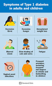
Associated symptoms are frequent urination, increased thirst, increased hunger, and weight loss.
Fluid loss causes dehydration and polydipsia.
Ketones are osmotically active and contribute to polyuria and dehydration.
Additional symptoms of type one diabetes may include increased hunger, fatigue, or blurred vision with swelling in the lens of the eye is due to chronically elevated blood sugar drawing in water.
Additional symptoms may include blurry vision, tiredness, and poor wound healing.
Symptoms typically develop rapidly.
Frequency is about 7.5% of diabetes cases.
Accounts for about 6% of all cases of diabetes in adults, and 80% of these cases are diagnosed before the patient is 20 years of age.
Accounts for approximately 6% of all cases of diabetes in adults of 18 years of age or greater in the US, and every percent of these cases are diagnosed before the patient is age 20.
Loss of beta cells is gradual with a substantial number remaining at clinical presentation and an ongoing decline after diagnosis of type one diabetes.
Amelioration of hyperglycemia after diagnosis allows partial recovery of beta cell insulin secretory function which can lead to a honeymoon, with relatively low exogenous insulin requirements.
The persistence of residual functioning beta cells, measured by means of C-peptide secretion is associated with improved glycemic control, a reduced risk of hypoglycemia, and a low incidence of micro vascular complications.
The cause of type 1 diabetes is believed to involve a combination of genetic and environmental factors.
Risk factors include having a family member with the condition.
The underlying abnormality involves an autoimmune destruction of the insulin-producing beta cells in the pancreas.
Diabetes is diagnosed by testing the level of sugar or glycated hemoglobin (HbA1C) in the blood.
Type 1 diabetes can be distinguished from type 2 by testing for the presence of autoantibodies.
Treatment with insulin is required for survival.
The disease requires a lifelong dependence on exogenous insulin.
A diabetic diet and exercise are important parts of management.
Hemoglobin A-1 C target of less than 7% is a chain by less than 20% of children with type one diabetes.
Tight glycemic control reduces the risk of diabetic microvascular complications in proportion to the degree of euglycemia that is reached.
Tight glycemic control extends to macrovascular disease with a 42% reduction in cardiovascular disease events over mean 17 year follow up.
Complications of relatively rapid onset include diabetic ketoacidosis and nonketotic hyperosmolar coma.
Long-term complications include heart disease, stroke, kidney failure, foot ulcers, neuropathy, and retinopathy.
In persons with diabetes type one, an elevated Lipo protein (a) level of greater than 50 milligrams per deciliter is a risk factor for the development of cardiovascular disease and albuminuria and is associated with poor glycemic control.
The use of aspirin for primary prevention of cardiovascular disease and patience with type one diabetes have yielded conflicting results.
Aspirin at low-dose use, as primary prevention is suggested for patients over the age of 50 and have other additional risk factors for premature atherosclerotic cardiovascular disease, hypertension, lipidemia, smoking, or chronic kidney disease.
A coronary artery calcium score of 100 or higher is associated with increase risk of cardiovascular disease among persons with and without type one diabetes.
Complications may arise from hypoglycemia caused by excessive dosing of insulin.
Type 1 diabetes makes up an estimated 5–10% of all diabetes cases.
Estimated 80,000 cases are diagnosed each year.
In the United States the number of people affected is estimated at 1-3 million.
Rates of disease vary widely geographically.
It typically begins in children and young adults.
Symptoms of type 1 diabetes include: polyuria, polydipsia, dry mouth, polyphagia, fatigue, and weight loss.
Polyuria developed when plasma glucose exceeds the kidneys ability to absorb it (greater than 180 mg/dL) so glucose is excreted in the urine, creating an osmotic effect that draws water into the urine.
Urinary excretion of glucose in addition to lipolysis from insulin deficiency causes weight loss.
Without insulin, glucose accumulates in the blood because muscle and fat cannot bring glucose transporters, specifically GLUT4, to the cell surface to allow glucose to enter.
When glucose is unavailable to muscle and other cells in the body for energy, the liver generates ketone bodies from fatty acids released from adipocytes, producing an alternative source of energy.
Ketone bodies are acidic and accumulation in the blood can cause, abdominal pain, nausea, vomiting, and Kussmaul breathing that occurs to exhale excess carbon dioxide, and reduce blood acidity.
It is often diagnosed when diabetic ketoacidosis occurs.
The signs and symptoms of diabetic ketoacidosis include: dry skin, rapid deep breathing, drowsiness, increased thirst, frequent urination, abdominal pain, and vomiting.
About 12 percent of people with type 1 diabetes have clinical depression.
About 6 percent of people with type 1 diabetes also have celiac disease.
In most cases patients with celiac disease and type one diabetes have are no digestive symptoms, mistakenly attribute problems to poor control of diabetes, gastroparesis or diabetic neuropathy.
In most cases, celiac disease is diagnosed after onset of type 1 diabetes.
Type one diabetes associated with celiac disease is associated with increased risk of complications, such as retinopathy, and mortality.
This association can be explained by shared genetic factors.
Patients with type 1 diabetes may experience dramatic and recurrent swings in glucose levels.
Unstable diabetes, labile diabetes or brittle diabetes results of swings of blood sugar that are irregular and unpredictable hyperglycemias, sometimes involving ketoacidosis, and sometimes serious hypoglycemias.
Unstable diabetes occurs in 1% to 2% of type I diabetics.
Type 1 diabetes is associated with alopecia areata, and is more common in the family members of people such hairless.
The cause of type 1 diabetes is unknown.
Theories on causes: genetic susceptibility, a diabetogenic trigger, and exposure to an antigen.
It involves multiple genes.
The risk of a child developing type 1 diabetes is:
about 5% if the father has it,
about 8% if a sibling has it,
and about 3% if the mother has it.
If one identical twin is affected there is about a 40% chance the other have type I diabetes.
Studies of heritability in association with type I diabetes is estimated at 80 to 86%.
More than 50 genes are associated with type 1 diabetes.
Such genes can be dominant, recessive, or somewhere in between.
IDDM1, is the strongest gene is located in the MHC Class II region on chromosome 6.
Variants of the IDDM1 gene increases the risk for decreased histocompatibility characteristic of type 1.
IDDM1 variants include DRB1 0401, DRB1 0402, DRB1 0405, DQA 0301, DQB1 0302 and DQB1 0201.
With Type 1 diabetes pancreatic β-cells are targeted and destroyed by the immune system, as a result of neo-natal mutations to the insulin gene (INS) which is responsible for mediating the production of the insulin in the pancreas.
The insulin gene is located on the short arm of chromosome 11.
In addition to chromosome 11, a genetic determinant of type 1 diabetes is a locus called the major histocompatibility complex (MHC) located on chromosome 6p21.
These above variants are common in North Americans of European ancestry and in Europeans.
The occurrence of type one diabetes varies by tenfold among individuals living in different areas of Europe.
The use of statins in type one diabetes, was shown to reduce cardiovascular events in a Swedish study (National Diabetes Register) but there are no randomized controlled studies to demonstrate positive effects of statins on atherosclerotic cardiovascular disease in patients with type one diabetes.
Some drugs destroy pancreatic islet cells: Streptozotocin (Zanosar), an antineoplastic agent, is selectively toxic to the beta cells of the pancreatic islets.
Trauma, pancreatitis, or tumors can also lead to loss of insulin production.
Checkpoint inhibitors inhibiting PD-1 and PD-L1), especially nivolumab and pembrolizumab have been reported to occasionally induce autoimmune diabetes.
The underlying pathophysiology in diabetes type 1 is a destruction of beta cells in the pancreas, regardless of causative entities.
The process that appears to be common to most risk factors is a type IV hypersensitivity autoimmune response towards beta cells, involving an expansion of autoreactive CD4+ T helper cells and CD8+ T cells, autoantibody-producing B cells and activation of the innate immune system.
The onset of autoimmune diabetes is accompanied by impaired ability to regulate the hormone glucagon.
Glucagon acts in antagonism with insulin to regulate blood sugar and metabolism.
Progressive beta cell destruction leads to dysfunction in the neighboring alpha cells which secrete glucagon.
Overproduction of glucagon after meals causes sharper hyperglycemia.
The failure to stimulate glucagon upon hypoglycemia prevents a glucagon-mediated rescue of glucose levels.
About 70% of patients with type one diabetes experience, some degree of hypoglycemia, each month.
Onset of type 1 diabetes is followed by an increase in glucagon secretion after meals.
Glucagon levels increases up to 37% during the first year of diagnosis, while c-peptide levels, indicative of islet-derived insulin, decline by up to 45%.
Insulin production falls as the immune system continues with progressive beta cell destruction, and islet-derived insulin continues to be replaced by exogenous insulin.
With time there is measurable alpha cell hypertrophy and hyperplasia in the early overt stage of the disease, leading to expanded alpha cell mass.
This, together with failing beta cell insulin secretion, accounts for rising glucagon levels that contribute to hyperglycemia.
It is suggested glucagon dysregulation is the primary cause of early stage hyperglycemia.
Staging: stages 1 and 2=are pre-symptomatic phases that are characterized by the development of autoimmunity and dysglycemia, respectively; stage 3 is defined as overt disease requiring exogenous insulin therapy.
Stage 2 type 1 diabetes is defined as the presence of two or more autoantibodies targeting insulin producing cells and impaired glycemic response is to glucose load, but otherwise normal metabolic disease, such as hemoglobin A1c, and no symptoms.
Hypoglycemia in type 1 diabetics is often a result of over-administered insulin therapy, though being in a fasting state, exercising without proper adjustment of insulin, sleep, and alcohol can also contribute.
The normal responses to hypoglycemia are impaired in type 1 diabetics.
Glucagon secretion increases with falling glucose levels, but normal glucagon response to hypoglycemia is blunted in type 1 diabetics and compared to healthy individuals experiencing an equal insulin-induced hypoglycemic trigger.
In type I diabetes beta cell glucose sensing and subsequent suppression of administered insulin secretion is absent, leading to islet hyperinsulinemia which inhibits glucagon release.
Recurrent episodes of hypoglycemia result in metabolic adjustments in the glucose sensing areas of the brain.
This brain hypoglycemic unawareness impairs sending of counter regulatory signals to the islets and adrenal cortex, and accounts for the lack of glucagon stimulation and epinephrine release that would normally stimulate glucose production from the liver, rescuing from severe hypoglycemia, coma, and death.
Autoimmune diabetes is characterized by a loss of islet specific sympathetic innervation.
Autoimmune diabetes are associated with an 80-90% reduction of islet sympathetic nerve endings.
The reduction of islet sympathetic nerve endings does not occur in type two diabetes.
Progressive autoimmune beta cell destruction causes the loss of protective factors to the islet sympathetic nerves.
This form of neuropathy is a hallmark of type 1 diabetes, and plays a part in the loss of glucagon rescue of severe hypoglycemia.
Hypertension is common in type one diabetes.
Hypertension is correlated with the duration of disease and the age of the population examined.
The prevalence of obesity in type one diabetes is greater than 36%.
Diagnostic criteria:
Fasting plasma glucose level at or above 7.0 mmol/l (126 mg/dl).
Plasma glucose at or above 11.1 mmol/l (200 mg/dl) two hours after a 75 g oral glucose load as in a glucose tolerance test.
Symptoms of hyperglycemia and casual plasma glucose at or above 11.1 mmol/l (200 mg/dl).
Glycated hemoglobin (hemoglobin A1C) at or above 48 mmol/mol (≥ 6.5 DCCT %).
About a quarter of people with new type 1 diabetes have developed some degree of diabetic ketoacidosis.
Diabetic ketoacidosis is a type of metabolic acidosis which is caused by high concentrations of ketone bodies, formed by the breakdown of fatty acids and the deamination of amino acids.
Diabetes may be diagnosed in a number of ways including:
Health screening, detection of hyperglycemia during other medical investigations, and secondary symptoms such as vision changes or unexplained fatigue, detected when a person suffers a problem that may be caused by diabetes, such as a heart attack, stroke, neuropathy, poor wound healing or a foot ulcer, certain eye problems, certain fungal infections, or delivering a baby with macrosomia or hypoglycemia.
Two fasting glucose measurements above 126 mg/dl (7.0 mmol/l) is considered diagnostic for diabetes.
In type 1 DM pancreatic beta cells in the islets of Langerhans are destroyed, decreasing endogenous insulin production.
Type 2 diabetes is characterized by insulin resistance, while type 1 diabetes is characterized by insulin deficiency, generally without insulin resistance.
The appearance of diabetes-related autoantibodies is able to predict the appearance of diabetes type 1 before any hyperglycemia arises.
The main autoantibodies are:
islet cell autoantibodies, insulin autoantibodies, autoantibodies targeting the 65-kDa isoform of glutamic acid decarboxylase (GAD), autoantibodies targeting the phosphatase-related IA-2 molecule, and zinc transporter autoantibodies (ZnT8).
All patients with autoantibodies progresses to diabetes type 1.
The risk of developing type I diabetes mellitus increases with the number of antibody types present.
The presence of three to four antibody types gives a risk of progressing to diabetes type 1 of 60–100%.
The latency time from the emergence of autoantibodies to clinically diagnosable diabetes can be a few months in infants and young children, to more than 10 years.
Immunofluorescence can detect Islet cell autoantibodies and other autoantibodies are measured with specific radiobinding assays.
The risk of heart failure is five times higher in women and three times higher in men with type one diabetes compared to controls.
Type 1 diabetes is not currently preventable.
Delaying onset of overt hypoglycemic disease in patients with stage one and two may be helpful for children, who stand to lose more than 14 years life expectancy is clinically diagnosed before age 10 years.
Cyclosporine A, is a immunosuppressive agent, that halts destruction of beta cells.
An anti-CD20 antibody, rituximab, inhibits B cells and has been shown to provoke C-peptide responses three months after diagnosis of type 1 diabetes, but long-term effects of this have not been reported.
Breastfeeding may decrease the risk of type 1 DM in later life.
Early introduction of gluten-containing cereals in the diet increases the risk of developing islet cell autoantibodies.
Children with antibodies to beta cell proteins without overt diabetes, and treated with niacinamide have less than half the diabetes onset incidence in a seven-year time span than did the general population.
Patients with type 1 diabetes and undiagnosed celiac disease have worse glycemic control and a higher prevalence of nephropathy and retinopathy.
Gluten-free diet, improves diabetes symptoms and appears to have a protective effect against developing long-term complications.
The evidence for the usefulness of low-carbohydrate dieting for people with type 1 diabetes is limited.
Patients with type 1 diabetes are advised to follow an individualized eating plan rather than a pre-decided one.
Treatment of type 1 diabetes is with the use of Insulin therapy.
Injections of insulin – via subcutaneous injection or using an insulin pump are necessary for those living with type 1 diabetes because it cannot be treated by diet and exercise alone.
Insulin dosage is adjusted based on food intake, blood glucose levels and physical activity.
Untreated type 1 diabetes can lead to diabetic ketoacidosis which can result in death, as a result cerebral edema.
Children are at a higher risk for cerebral edema than adults.
Ketoacidosis is the most common cause of death in pediatric diabetes.
Treatment focuses on lowering blood sugar or glucose to the near normal range.
The initial phase of treatment includes stabilization of glucose levels and education of patient/family about monitoring of blood glucose, techniques for insulin injection, dose calculations, nutritional principles, interpretation of glucose levels, and management of hypoglycemia.
The combination of exogenous basal and bolus insulin delivery by injections and self monitoring of blood glucose 4-6 times a day is most common.
Subsequent utilization of insulin pumps, continuous glucose monitoring, advanced insulin formulations and other technological advances are introduced.
Continuous glucose monitoring eliminates the need for frequent and painful fingersticks.
Guidelines recommend glycemic targets include time in range over a 24 hour period that the blood glucose level is in the reference range of 70 to 180 mg/dL of more than 70%, amounting to approximately 17 hours per day.
The ultimate goal of normalizing glucose levels is to avoid complications: peripheral neuropathy, cardiovascular system, retinopathy.
This level of control over a prolonged period of time can be varied by a target HbA1c level of less than 7.5%.
There are four main types of insulin: rapid acting insulin, short-acting insulin, intermediate-acting insulin, and long-acting insulin.
The rapid acting insulin is administered as a bolus dosage, and its
action onsets in 15 minutes with peak actions in 30 to 90 minutes.
Short acting insulin action onsets within 30 minutes with the peak action around 2 to 4 hours.
Intermediate acting insulin action onsets within one to two hours with peak action of four to 10 hours.
Long-acting insulin is usually given at once per day, and its action onset is roughly 1 to 2 hours with a sustained action of up to 24 hours.
Some insulins are biosynthetic products produced using genetic recombination techniques.
Previously cattle or pig insulins were used.
Hypoglycemia commonly occurs in people with diabetes, usually the result of a mismatch in the balance among insulin, food and physical activity.
Hypoglycemia symptoms include:
excess sweating, excessive hunger, fainting, fatigue, lightheadedness and shakiness.
Mild hypoglycemia is self treated for eating or drinking something with a high sugar content.
Severe hypo glycemia can lead to unconsciousness and are treated with intravenous glucose or injections with glucagon.
Artificial pancreas management is promising.
In some cases, a pancreas transplant can restore proper glucose regulation, and is generally only used with or some time after a kidney transplant.
Pancreas transplants alone may be beneficial in people with extremely labile type 1 diabetes.
Islet cell transplantation may be an option for some people with type 1 diabetes.
Restoring the function of islets can lead to physiological glycemic control without an increased risk of hypoglycemia.
Beta cell replacement with use of islet or pancreas transplantation reduces, or eliminates the need for insulin therapy, which decreases the risk of severe hypoglycemia caused by insulin.
Beta cell replacement, however, is limited by organ availability and variable islet quality.
The need for multiple transplants from multiple donors to achieve acceptable. linical outcomes limits the usefulness of islet transplantation.
Complications of diabetes type1 :
cardiovascular disease, diabetic neuropathy, and diabetic retinopathy, among others.
Women with type 1 DM have a 40% higher risk of death as compared to men with type 1 DM.
The life expectancy of an individual with type 1 diabetes is 11 years less for men and 13 years less for women.
Patients with type 1 diabetes are higher risk for other autoimmune diseases: autoimmune thyroid disease, celiac disease, rheumatoid arthritis, and lupus.
There is an increased rate of urinary tract infection due to bladder dysfunction.
Males with diabetes are subject to problems with erections and ejaculation.
Sexual problems are common in women who have diabetes: reduced sensation in the genitals, vaginal dryness, difficulty/inability to orgasm, pain during sex, and decreased libido.
Women with type 1 diabetes show a higher rate of polycystic ovarian syndrome (PCOS).
Over 18,000 youths are diagnosed with Type 1 diabetes every year.
Vaccines are being looked at to treat or prevent type 1 diabetes by inducing immune tolerance to insulin or pancreatic beta cells.
Autoimmune destruction of beta cells in type I diabetes begins before the onset of hyperglycemia and measurement of C- peptide responses at the time of diagnosis demonstrates that some beta cell function at this stage is retained.
Residual beta cell function in type I diabetes is associated with reductions of severe hypoglycemic episodes and its complications (The Diabetes Control and Complications Trial Research Group).
Patients with mild Type I diabetes may initially have enough beta cell function to prevent acidosis, but later in life insulin dependence will result as the beta cell function diminishes.
Mild type I DM may make up 15% of diabetes patients, known as latent autoimmune diabetes of adulthood.
Type I diabetes is more common in Scandinavian countries and becomes less frequent closer to the equator.
The incidence of type I diabetes increases for populations that emigrate to the northern hemisphere.
Fewer than 10% of patients with type I DM have no evidence for pancreatic B cell autoimmunity explaining absent insulin and ketoacidosis-this type of diabetes is known as idiopathic type I diabetes or type IB diabetes.
Idiopathic type I diabetes is most commonly seen in individuals of Asian or African origin.
Average life expectancy of a person who has diabetes is 15 years less than a person who does not have the disease.
The use of a closed end loop system that automates insulin delivery in a glucose responsive manner (automated insulin delivery system or artificial pancreas) has the potential to improve glycemic outcomes quality-of-life in children than the use of a sensor augmented insulin pump (Breton MD).
A hybrid closed-loop system significantly improves glycemic control in very young children with type one diabetes, without increasing periods of hypoglycemia.
A hybrid closed-loop system, which is also called an artificial pancreas, in which an algorithm automatically adjusts insulin delivery on the basis of real time sensory glucose levels.
An open loop system improves the time in optimal glucose management for type one diabetics.
In youths with new onset type one diabetes intensive glucose control for 24 months does not prevent the decline in residual C-peptide secretion.
The use of continuous glucose monitors in children, with or without pump use, has shown substantial benefits for glycemic control.
Continuous glucose monitoring compared with self monitoring of blood glucose results and lowered hemoglobin A1c levels, lower rates of hypoglycemia, reduced incidence of diabetic ketoacidosis,and improved psychosocial outcomes and quality of life.
In young people with newly diagnosed type one diabetes, intensive diabetes management, including automated insulin delivery, achieved, excellent glucose control, but did not affect the decline in pancreatic C-peptide secretion at 52 weeks (McVean J).
Glarine, determir, and the gludec are the most common preparations of basal insulin used in the treatment of children and adolescents with type one diabetes.
NPH insulin is less expensive and readily available.
However, it requires two daily injections whereas one daily injection is usually sufficient with the above insulins.
Degludic and Glarine have a half-life of approximately 25 hours, low peak to trough ratio, and nearly flat action profile with less risk for hypoglycemia.
Teplizumab, a monoclonal antibody can delay the onset of clinically diagnosed stage 3 type 1diabetes by a median of about two years.
Teplizumab is the first medication approved to slow the progression of type1 diabetes and need for insulin therapy.
Two 12 day courses of teplizumab in children in adolescents, with newly diagnosed type one diabetes showed benefit with respect to the primary endpoint of preservation of beta cell function, but not but did not show significant differences with respect to secondary endpoints.
Immunomodulating agents have been shown to preserve some beta cell function in recent onset type one diabetes including: cyclosporine, teplizumab, abatacept, alefacept, rituximab, golimumab, low-dose, antithymocyte globulin, and the combination of anti-IL – 21 monoclonal antibody with liraglutide.
Verapamil in patients with recent onset type one diabetes, modestly preserves endogenous insulin secretion.
Verapamil treatment resulted in a 30% increase in C-peptide secretion compared with placebo and delayed the expected decline of C-peptide production from 3 to 6 months after diagnosis early onset of type one diabetes.
In patients with type 1 diabetes of recent onset, daily treatment with baricitinib over 48 weeks preserved beta cell function as estimated by the mixed meal stimulated main C-peptide level.