 Transbronchial biopsy (TBB) is performed to diagnose focal and diffuse lung diseases.
Transbronchial biopsy has lower morbidity and mortality than open lung biopsy.
Transbronchial biopsy has two main rare complications: pneumothorax and pulmonary bleeding.
Endobronchial ultrasonography (EBUS)-guided needle aspiration can be used for mediastinal lymph nodes if these are present and can replace transbronchial biopsy, especially for suspected sarcoidosis.
Indications for transbronchial biopsy:
Neoplastic disease
Suspected sarcoidosis
Hypersensitivity pneumonitis
Interstitial lung disease [
Pulmonary infection
Unclear lung disease
Lung transplantation
Lung nodules and masses.
TBB’s diagnostic accuracy increases when the nodule is larger than 2 cm, when the presence of a bronchus leading to the nodule is found on CT of the chest. and when the tissue is repeatedly sampled.
The yield of transbronchial biopsy is also high in bronchoalveolar carcinoma and lymphangitic spread of the tumor.
The diagnostic yield of TBB is increased to 73% by combining flexible bronchoscopy with CT guidance in a dedicated low-dose protocol.
Transbronchial biopsy has not always been found to be reliable for heterogeneous lung diseases such as usual interstitial pneumonia.
Transbronchial biopsy has high diagnostic yield for sarcoidosis: Sensitivity ranges from 50% to 85% in stage 1 disease and is higher if the parenchyma is involved.
Transbronchial biopsy is also highly sensitive in diagnosing pulmonary alveolar proteinosis, Langerhans cell histiocytosis, eosinophilic pneumonia, lipoid pneumonia, drug-induced pneumonitis, and miscellaneous lung disease.
Transbronchial biopsy is indicated in the diagnosis of pulmonary infections in the immunocompromised patient.
Transbronchial biopsy increases the diagnostic yield for Pneumocystis jiroveci pneumonia.
It is used in the diagnosis of allograft rejection in lung transplantation.
Absolute contraindications for transbronchial biopsy:
Medical instability
Severe hypoxia
Status asthmaticus
Lack of patient cooperation
Malignant arrhythmia
Massive hemoptysis
Uncorrectable bleeding diathesis
Relative contraindications include:
Thrombocytopenia –
Uremia – biopsies may be done carefully, preferably soon after dialysis or administration of desmopressin (DDAVP) and cryoprecipitate
Transbronchial biopsy risks should be weighed against the risk of developing tension pneumothorax.
It is suggested TBB should be performed with caution in patients with elevated pulmonary arterial pressures.
It is recommended that oxygen supplementation to keep the blood oxygen saturation above 90% and thereby minimize the risk of cardiac arrhythmia during and after bronchoscopy.
Certain antiplatelet and anticoagulant drugs are discontinued beforehand.
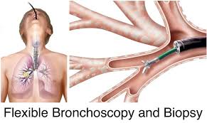
The flexible bronchoscope is wedged in the segmental bronchus of interest, and the biopsy forceps is passed through the working channel of the bronchoscope.
The number of biopsy specimens required for optimal diagnostic yield has been reported to be four to 10.
It is recommended that at least five samples should be obtained in cases where endobronchial tumor is visible and that at least five or six samples should be obtained in cases of interstitial lung disease.
The ideal transbronchial biopsy specimen consists of four to six samples, with at least one sample containing full-thickness bronchial mucosa and some alveolar parenchyma.
The use of fluoroscopy during transbronchial biopsy has been shown to increase the diagnostic yield in focal lesions, but has not been found to provide a comparable benefit in diffuse lung diseases such as sarcoidosis.
The tissue samples obtained by means of transbronchial biopsy forceps are about 3 mm.
TBB is less useful in diagnosing heterogeneous lung diseases such as idiopathic interstitial fibrosis.
Combining suction catheter aspiration and forceps biopsy results in a higher diagnostic yield than use of either method by itself.
The use of fluoroscopy equipment may increase the diagnostic yield and provide a statistically insignificant decrease in pneumothoraces.
Computed tomography (CT) of the chest is usually performed before bronchoscopy helps predict the yield of transbronchial biopsy on the basis of the anatomic distribution and appearance of any abnormalities.
Peribronchovascular and central abnormalities are much more amenable to specific diagnosis by transbronchial biopsy than peripheral or nonsegmental disease is.
Transbronchial biopsy is often diagnostic in patients in whom the CT findings are those of centrilobular nodules of ground-glass attenuation.
TBB must be for monitored for blood pressure, oxygen saturation, heart rate, respiratory rate, and possibly end-tidal CO2 and equipment includes A flexible bronchoscope, a light source, video monitoring equipment, a biopsy forceps, specimen containers, equipment for cardiopulmonary resuscitation, a suction apparatus, and supplemental oxygen.
Biplane fluoroscopy equipment is necessary for accurate localization of lesions.
Transbronchial biopsy is usually performed with the patient in the supine position.
Typically, topical anesthesia with lidocaine is applied to the nasal mucosa, the oropharynx, and larynx.
The patient under conscious sedation induced by intravenous (IV) narcotics and benzodiazepines.
Bronchoscopic biopsies conventionally performed with forceps, which can result in small specimens and poor specimen quality due to crush artifact.
General anesthesia may be required if rigid bronchoscopy is performed.
Midazolam is the benzodiazepine of choice for short-term sedation because it has the highest lipid solubility, the fastest onset of action of 1-5 minutes, and the shortest duration of action at 1-2 hours.
Fentanyl is the narcotic most frequently used for conscious sedation, being 600 times more lipid-soluble than morphine and thus is more readily taken up into the central nervous system.
Propofol is increasingly used.
Technical advances are expected to increase diagnostic yield, avoiding transthoracic needle aspiration more invasive video-assisted thoracoscopic surgery (VATS) in the operating room.
Endobronchial ultrasonography (EBUS) helps guide the bronchopist to the peripheral lesion.
Electromagnetic Navigational Bronchoscopy (ENB)is a minimally invasive approach to guide bronchoscopic biopsies or needle aspiration to small pulmonary nodules.
ENB is particularly useful in the setting of lung nodules; the detection of early lung cancer is important to allow early successful treatment.
In ENB, a computer system converts images from a patient’s CT scan to construct a 3D road map for the purpose of guiding bronchoscopy, particularly in the endobronchial tree.
It can guideadvancement of needles, forceps, and brushes into small pulmonary nodules.
ENB achieves a diagnostic yield of 67-74% with complication of pneumothorax rates of 2.2-3.5%.
Electromagnetic navigational bronchoscopy can also serve to place fiducial markers, which are useful for guidance of stereotactic radiotherapy or VATS.
With ENB it is possible to avoid thoracic surgery for benign nodules.
Nodule location changes with respiration during bronchoscopy and is not captured with an end-inspiratory CT.
Transbronchial lung biopsy performed with a 1.1 mm cryoprobe had a significantly higher diagnostic yield compared with 2 mm forceps in patients with lung nodules or masses, lung transplant, and diffuse parental lung disease.
The major complications of transbronchial biopsy are pneumothorax and bleeding: Pneumothorax occurs in 1-6% of patients undergoing transbronchial biopsy.
Chest radiographs are not routinely necessary after the procedure.
Significant bleeding occurs in 2-9% of procedures.
Unusual complications include mediastinal and subcutaneous emphysema.