 1122
1122
A muscular organ in the mouth that manipulates food for mastication, and is used in the act of swallowing.
It is important in the digestive system.
It is the primary organ of taste in the gustatory system.
Its upper surface is covered by taste buds housed in numerous lingual papillae.
In health, the dorsal surface of the tongue is covered in tuft-like projections called lingual papillae. (some of which are associated with taste buds.
Papillae which give the tongue an irregular surface texture and a white-pink color.
It is sensitive tissue, kept moist by saliva, and serves as a natural means of cleaning the teeth.
It is richly supplied with nerves and blood vessels.
One of its major functions is the enabling of speech.
It is divided into two parts, an oral part at the front and a pharyngeal part at the back.
It is divided into anterior and posterior parts by the terminal sulcus which is a V-shaped groove.
The apex of the terminal sulcus is marked by a blind foramen, the foramen cecum, which is a remnant of the median thyroid diverticulum in early embryonic development.
The anterior oral part of the ton is the visible part situated at the front.
The anterior tongue makes up roughly two-thirds the length of the tongue.
The posterior pharyngeal part is the part closest to the throat, comprises roughly one-third of its length.
The left and right sides of the tongue is separated along most of its length by fibrous tissue, the lingual septum.
The lingual septum results in a groove, the median sulcus on the tongue’s surface.
The tongue has two groups of muscles.
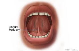
The tongue is attached to the floor of the mouth by a ligamentous band called the frenum and this gives it great mobility for the manipulation of food, and speech.
There are four intrinsic muscles alter the shape of the tongue and are not attached to bone.
The four paired extrinsic muscles change the position of the tongue and are anchored to bone.
The range of manipulation of then tongue is optimally controlled by the action of several muscles and limited in its external range by the stretch of the frenum.
The tongue’s two sets of muscles, are four intrinsic muscles that originate in the tongue and are involved with its shaping, and four extrinsic muscles originating in bone that are involved with its movement.
It forms part of the floor of the oral cavity.
The anterior tongue is, at its apex thin and narrow.
The anterior tongue is directed forward against the lingual surfaces of the lower incisor teeth.
The posterior part of the tongue is, at its root, directed backward, and connected:
with the hyoid bone by the hyoglossi and genioglossi muscles
the hyoglossal membrane,
with the epiglottis by three glossoepiglottic folds of mucous membrane,
with the soft palate by the glossopalatine arches,
with the pharynx by the superior pharyngeal constrictor muscle and the mucous membrane.
The tongue forms the anterior wall of the oropharynx.
The average length of the human tongue from the oropharynx to the tip is 10 cm.
The average weight of the human tongue from adult males is 70g and for adult females 60g.
Apiical sounds are made with the tip of the tongue, while laminal sounds are generated from the tongue blade, behind the tip.
The upper surface of the tongue is referred to as the dorsum.
The dorsum of the tongue is divided by a groove into symmetrical halves by the median sulcus.
At about 2.5 cm from the root of the tongue, the foramen cecum marks the end of this division and the beginning of the terminal sulcus.
The thyroglossal duct attaches at the foramen cecum and is formed during the descent of the thyroid diverticulum in embryonic development.
The pharyngeal part of the tongue is supplied by the glossopharyngeal nerve.
The oral part of the tongue is supplied by the lingual nerve, which is a branch of the mandibular branch (V3) of the trigeminal nerve for somatosensory perception and by the chorda tympani, a branch of the facial nerve, for taste perception.
Embryologically both parts of the tongue develop from different pharyngeal arches.
The four extrinsic muscles of the tongue originate from bone and extend to the tongue.
They are the genioglossus, the hyoglossus, the styloglossus, and the palatoglossus.
Their main functions of the extrinsic muscles of the tongue are to change the tongue’s position allowing for protrusion, retraction, and side-to-side movement.
The genioglossus is the only muscle that propels the tongue forward, and arises from the mandible.
Arising from the hyoid bone, the hyoglossus muscle retracts and depresses the tongue.
The chondroglossus muscle is often included with the hyoglossus muscle
The styloglossus muscle arises from the styloid process of the temporal bone.
The styloglossus muscle draws the sides of the tongue up to create a trough for swallowing.
The palatoglossus muscle arises from the palatine aponeurosis, and depresses the soft palate.
The palatoglossus muscle moves the palatoglossal fold towards the midline, and elevates the back of the tongue during swallowing.
There are 4 paired intrinsic muscles of the tongue, originating and inserting within the tongue, running along its length.
The intrinsic tongue muscles are the superior longitudinal muscle, the inferior longitudinal muscle, the vertical muscle, and the transverse muscle.
The intrinsic muscles of the tongue alter its shape
by lengthening, shortening, curling and uncurling its apex and edges.
The rolling, flattening and rounding of the tongue surface is accomplished by the intrinsic muscles.
The shaping of the tongue facilitates speech, swallowing, and eating.
The superior longitudinal muscle runs along the upper surface of the tongue under the mucous membrane.
The superior longitudinal muscle elevates, and assists in retraction of, or deviates the tip of the tongue.
The superior longitudinal muscle originates near the epiglottis, at the hyoid bone, from the median fibrous septum.
The inferior longitudinal muscle lines the sides of the tongue, and is joined to the styloglossus muscle.
The vertical muscle is located in the middle of the tongue.
The vertical muscle joins the superior and inferior longitudinal muscles.
The transverse muscle divides the tongue at the middle and runs along the sides, attached to the mucous membranes.
The lingual artery, a branch of the external carotid artery, is the primary blood supply to the tongue,and the floor of the mouth.
A secondary blood supply to the tongue is from the tonsillar branch of the facial artery and the ascending pharyngeal artery.
Lighting the lingual artery helps to stop severe hemorrhage from the tongue.
Blood is drained into the internal jugular vein via the lingual veins.
Tongue innervation consists of motor fibers, special sensory fibers for taste, and general sensory fibers for sensation.
Motor supply for all intrinsic and extrinsic muscles of the tongue is supplied by efferent motor nerve fibers from the hypoglossal nerve (CN XII), with the exception of the palatoglossus.
The palatoglossus muscle is innervated by the vagus nerve (CN X).
The innervation of taste and sensation is different for the anterior and posterior part of the tongue
The anterior tongue id derived from pharyngeal arch 1.
The posterior tongue is derived from pharyngeal arches 3 and 4.
Innervation of the anterior two thirds of tongue:
Taste: chorda tympani branch of the facial nerve (CN VII) via special visceral afferent fibers.
Sensation: lingual branch of the mandibular (V3) division of the trigeminal nerve (CN V) via general visceral afferent fibers
Innervation of Posterior one third of tongue:
Taste and sensation: glossopharyngeal nerve (CN IX) via a mixture of special and general visceral afferent fibers
Base of tongue:
Taste and sensation: internal branch of the superior laryngeal nerve (itself a branch of the vagus nerve, CN X
The upper surface of the tongue is covered in masticatory mucosa.
Masticatory mucosa is a type of oral mucosa which is of keratinized stratified squamous epithelium.
Embedded in this are numerous
Papillae are embedded in the mucosa that house the taste buds and their taste receptors.
The tongue papillae consist of a number of types: filiform, fungiform, vallate, and foliate.
The filiform papillae are not associated with any taste buds, while the other types are.
The lingual papillae covers the dorsal side of the tongue, while the ventral surface is stratified squamous non-keratinized epithelium which is smooth.
Tastants are chemicals that stimulate taste receptor cells.
When tastant is dissolved in saliva, it can make contact with the plasma membrane of the gustatory hairs.
The gustatory hairs are the sites of taste transduction.
It is equipped with many taste buds on its dorsal surface, and each taste bud is equipped with taste receptor cells that can sense particular classes of tastes.
Taste receptor cells are distinct and detect substances that are sweet, bitter, salty, sour, spicy, or taste of umami.
Umami receptor cells are the least understood.
The tongue is used for crushing food against the hard palate, during mastication and manipulation of food for softening prior to swallowing, and is therefore is an important accessory organ in the digestive system.
Because the epithelium on the tongue’s upper surface is keratinized, it can grind against the hard palate without being itself damaged or irritated.
The intrinsic muscles of the tongue allow for the shaping of the tongue which facilitates speech.
The tongue has a role in physical intimacy and sexuality.
In an congenital disorder of the tongue ankyloglossia, the tongue is tied to the floor of the mouth by a very short and thickened frenulum and this affects speech, eating, and swallowing.
Tongue pathologies include: glossitis, geographic tongue, median rhomboid glossitis, burning mouth syndrome, oral hairy leukoplakia, oral candidiasis and black hairy tongue.
Squamous cell carcinomas can affect the tongue.
Food debris, desquamated epithelial cells and bacteria often form a visible tongue coating, which can be a major contributing factor in bad breath.
The sublingual region underneath the front of the tongue is a location for the administration of medications.
The sublingual region of the tongue has an oral mucosa that is very thin and has underlying plexus of veins, and allows for the speedy application of medication into the blood, bypassing the gastrointestinal tract.