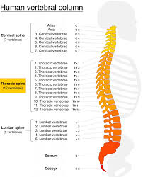
Thoracic vertebrae compose the middle segment of the vertebral column, between the cervical vertebrae and the lumbar vertebrae.
There are twelve thoracic vertebrae and they are intermediate in size between the cervical and lumbar vertebrae.
Thoracic vertebrae increase in size going towards the lumbar vertebrae, with the lower ones being much larger than the upper.
Thoracic vertebrae have facets on the sides of the bodies for articulation with the heads of the ribs, as well as facets on the transverse processes of all, except the eleventh and twelfth, for articulation with the tubercles of the ribs.
Thoracic vertebrae are numbered T1–T12, with the first one (T1) located closest to the skull and the others going down the spine toward the lumbar region.
Characteristics of the second through eighth thoracic vertebrae.
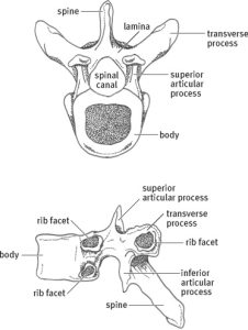
The bodies in the middle of the thoracic region are heart-shaped: as broad in the anteroposterior as in the transverse direction.
At the ends of the thoracic region the bodies of the thoracic vertebra resemble respectively those of the cervical and lumbar vertebrae.
At the ends of the thoracic region the vertebrae are slightly thicker behind than in front, flat above and below, convex from side to side in front, deeply concave behind, and slightly constricted laterally and in front.
The mid bodies present, on either side, two costal demi-facets, one above, near the root of the pedicle, the other below, in front of the inferior vertebral notch.
The facets are covered with cartilage in and, when the vertebrae are articulated with one another, form, with the intervening intervertebral fibrocartilages, oval surfaces for the reception of the heads of the ribs.
The pedicles are directed backward and slightly upward, and the inferior vertebral notches are of large size.
The laminae are broad, thick, and overlap those of subjacent vertebrae and connect with the pedicles to surround and protect the spinal cord.
The intervertebral foramen is small, and circular, with two at each intervertebral level, one for the right and one for the left exiting nerve roots.
The vertebral foramen is the large opening posterior to the vertebral body also known as the spinal canal, containing the spinal cord at the thoracic level.
The spinal canal protects the spinal cord.
The thoracic spinous processes are long, triangular and directed obliquely downward, arising from the lamina and ending in a tuberculated extremity.
These spinous processes overlap from the fifth to the eighth thoracic vertebrae, but are less oblique in direction above and below.
The superior articular processes are thin plates of bone projecting upward from the junctions of the pedicles and laminae.
The articular facets are practically flat, and are directed backward and a little lateralward and upward.
The inferior articular processes are fused to the laminae, and project slightly beyond their lower borders.
The inferior articular process facets are directed forward and a little medialward and downward.
The transverse processes arise from the arch behind the superior articular processes and pedicles.
The transverse processes are thick, strong, and directed obliquely backward and lateralward, and each ends in a clubbed extremity, on the front of which is a small, concave surface, for articulation with the tubercle of a rib.
The first and ninth through twelfth vertebrae contain certain peculiarities.
The first thoracic vertebra has, on either side of the body, an entire articular facet for the head of the first rib, and a demi-facet for the upper half of the head of the second rib.
The body of the first thoracic vertebra is like that of a cervical vertebra, being broad, concave, and lipped on either side.
The superior articular surfaces are directed upward and backward; the spinous process is thick, long, and almost horizontal.
The transverse processes are long, and the upper vertebral notches are deeper than those of the other thoracic vertebrae.
The thoracic spinal nerve 2 (T2) passes out underneath the second thoracic vertebrae.
The second thoracic vertebra is larger than the first thoracic vertebra.
The thoracic spinal nerve 3 (T3) passes out underneath the third thoracic vertebra (T3).
The fourth thoracic (T4) vertebra, together with the fifth, is at the same level as the sternal angle.
The thoracic spinal nerve 4 (T4) passes out underneath it.
As noted the fifth thoracic vertebra, together with the fourth, are at the same level as the sternal angle.
The human trachea divides into two main bronchi at the level of the 5th thoracic vertebra, but may also end higher or lower, depending on breathing.
The thoracic spinal nerve 5 (T5) passes out underneath it.
The thoracic spinal nerve 6 (T6) passes out underneath it.
The thoracic spinal nerve 7 (T7) passes out underneath it.
The eighth thoracic vertebra is, together with the ninth thoracic vertebra, at the same level as the xiphisternum.
The thoracic spinal nerve 8 (T8) passes out underneath it.
The ninth thoracic vertebra (T9) may have no demi-facets below.
The thoracic spinal nerve 9 (T9) passes out underneath it.
The tenth thoracic vertebra (T10) has an entire articular facet on either side, which is placed partly on the lateral surface of the pedicle.
The thoracic spinal nerve 10 (T10) passes out underneath it.
The eleventh thoracic vertebra (T11)
body approaches in its form and size to that of the lumbar vertebrae.
The articular facets for the heads of the ribs are of medium size, and placed chiefly on the pedicles, which are thicker and stronger in this and the next vertebra than in any other part of the thoracic region.
The spinous process is short, and nearly horizontal in direction.
The transverse processes are very short, tuberculated at their extremities, and do not have articular facets.
The thoracic spinal nerve 11 (T11) passes out underneath it.
Twelfth thoracic vertebra (T12) has the same general characteristics as the eleventh, but may be distinguished from it by its inferior articular surfaces being convex and directed lateralward, like those of the lumbar vertebrae.
The general form of the T12 body, laminae, and spinous process resembles the lumbar vertebrae; and by each transverse process being subdivided into three elevations, the superior, inferior, and lateral tubercles:
The superior and inferior correspond to the mammillary and accessory processes of the lumbar vertebrae.
The thoracic spinal nerve 12 (T12) passes out underneath it.