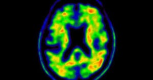
 A tau positron emission tomography (PET) scan is a molecular imaging technique that uses radiolabeled tracers to visualize and quantify the distribution of aggregated tau protein in the brain.
Tau is a microtubule-associated protein that, when abnormally phosphorylated and aggregated, forms neurofibrillary tangles—a pathological hallmark of Alzheimer disease (AD) and other tauopathies.
Tau PET imaging enables detection of these tangles, providing a biomarker for tau pathology that correlates closely with neurodegeneration and clinical symptoms in AD.
The most widely used tau PET tracer is [18F]flortaucipir, which binds to paired helical filament tau found in advanced stages (V–VI) of AD.
A positive tau PET scan reflects the presence of significant neocortical tau pathology, which is strongly associated with cognitive impairment and dementia.
Tau PET’s clinical utility is highest in distinguishing AD from non-AD dementias, especially in symptomatic individuals, and it is increasingly used as a biomarker in clinical trials of disease-modifying therapies.