 The striatum, or corpus striatum is a cluster of neurons in the subcortical basal ganglia of the forebrain.
The striatum is a part of the brain that plays a crucial role in motor control, motivation, reward, and reinforcement learning.
It is primarily involved in the coordination and execution of voluntary movements.
The striatum controls posture and movement.
The striatum receives input from various regions of the brain and integrates information to generate appropriate motor responses.
It also plays a role in the regulation of emotions, decision-making, and the processing of rewards and punishments.
Dysfunction in the striatum has been associated with various neurological and psychiatric disorders, including Parkinson’s disease, Huntington’s disease, and addiction.
The striatum is a critical component of the motor and reward systems.
It receives glutamatergic and dopaminergic inputs from different sources.
The striatum serves as the primary input to the rest of the basal ganglia.
It coordinates multiple aspects of cognition, including both motor and action planning, decision-making, motivation, reinforcement, and reward perception.
The striatum is made up of the caudate nucleus and the lentiform nucleus.
The lentiform nucleus is made up of the larger putamen, and the smaller globus pallidus.
It is common practice, however, to implicitly exclude the globus pallidus when referring to striatal structures.
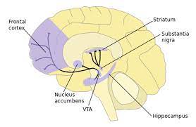
The striatum is divided into a ventral striatum, and a dorsal striatum, based upon function and connections.
The ventral striatum consists of the nucleus accumbens and the olfactory tubercle.
The dorsal striatum consists of the caudate nucleus and the putamen.
A white matter, nerve tract the internal capsule in the dorsal striatum separates the caudate nucleus and the putamen.
Anatomically, the term striatum describes its striped appearance of grey-and-white matter.
The striatum is the largest structure of the basal ganglia.
The nucleus accumbens is made up of the nucleus accumbens core and the nucleus accumbens shell, which differ by neural populations.
The olfactory tubercle receives input from the olfactory bulb but has not been shown to play a role in processing smell.
The ventral striatum is associated with the limbic system and has been implicated as a vital part of the circuitry for decision making and reward-related behavior.
Types of cells in the striatum include:
Medium spiny neurons (MSNs) are the principal neurons of the striatum, compriseing 95% of the total neuronal population and they are GABAergic and classified as inhibitory neurons.
Medium spiny neurons have two characteristic types: D1-type MSNs and D2-type MSNs.
Approximately 40% of striatal MSNs expressing both DRD1 and DRD2 mRNA.
Cholinergic interneurons release acetylcholine, which important effects in the striatum.
These interneurons respond to environmental stimuli with stereotyped responses that are temporally aligned with the responses of dopaminergic neurons of the substantia nigra.
The large aspiny cholinergic interneurons themselves are affected by dopamine receptors.
Dopamine also directly controls communication between cholinergic interneurons.
There are many types of GABAergic interneurons, which participate in feedforward inhibition of principal neurons.
GABAergic interneurons that express tyrosine hydroxylase, somatostatin, nitric oxide synthase and neuropeptide-y.
The striatum has its own internal microcircuitry.
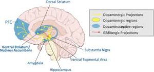
The ventral striatum receives direct input from multiple regions in the cerebral cortex and limbic structures such as the amygdala, thalamus, and hippocampus, entorhinal cortex and the inferior temporal gyrus.
The corpus striatum’s primary input is to the basal ganglia system.
Additionally, the mesolimbic pathway projects from the ventral tegmental area to the nucleus accumbens of the ventral striatum.
The nigrostriatal connection arises from the neurons of the substantia nigra pars compacta and is glutamatergic.
The striatum receives additional afferents from other elements of the basal ganglia such as the subthalamic nucleus (glutamatergic) or the external globus pallidus (GABAergic).
Striatal outputs from both the dorsal and ventral components are primarily composed of medium spiny neurons( MSNs): indirect MSNs that express D2-like receptors and direct MSNs that express D1-like receptors.
The main nucleus of the basal ganglia is the striatum.
The stratum projects directly to the globus pallidus via a pathway of striatopallidal fibers.
The striato-pallidal pathway has a whitish appearance due to the myelinated fibers.
The striato-pallidal pathway comprises successively the external globus pallidus (GPe), the internal globus pallidus (GPi), the pars compacta of the substantia nigra (SNc), and the pars reticulata of substantia nigra (SNr).
The neurons of this projection are inhibited by GABAergic synapses from the dorsal striatum.
Deep penetrating striate arteries supply blood to the striatum.
The ventral striatum, and the nucleus accumbens in particular, primarily mediate reward, cognition, reinforcement, and motivational salience.
The dorsal striatum primarily mediates cognition involving motor function, certain executive functions and stimulus-response learning.
Some small degree of overlap exists as the dorsal striatum is also a component of the reward system that, along with the nucleus accumbens core, mediates the encoding of new motor programs associated with future reward acquisition.
The striatum is activated by stimuli associated with reward, but also by aversive, novel, unexpected, or intense stimuli, and cues associated with such events.
fMRI evidence suggests that the common property linking these stimuli, to which the striatum is reacting, is salience under the conditions of presentation.
The interplay between the striatum and the prefrontal cortex is relevant for behavior, particularly adolescent development.
Parkinson’s disease results in loss of dopaminergic innervation to the dorsal striatum, and other basal ganglia.
Atrophy of the striatum is also involved in Huntington’s disease, chorea, choreoathetosis, and dyskinesias.
Addiction is a disorder of the brain’s reward system.
Addiction arises through the overexpression of DeltaFosB (ΔFosB), a transcription factor, in the D1-type medium spiny neurons of the ventral striatum.
ΔFosB is an inducible gene which is increasingly expressed in the nucleus accumbens as a result of repeatedly overdosing on an addictive drug or overexposure to other addictive stimuli.
An association between striatal expression of variants of the PDE10A gene and some bipolar I disorder patients.
Variants of other genes, DISC1 and GNAS, have been associated with bipolar II disorder.
Autism spectrum disorder (ASD) is characterized by cognitive inflexibility and poor understanding of social systems, which originates in defects in the pre-frontal cortex as well as the striatal circuits.
The defects in the striatum seem to specifically contribute to the motor, social and communication impairments seen in ASD patients.
Dysfunction in the ventral striatum can lead to disorders, most notably, depression and obsessive-compulsive disorder.
Because of its involvement in reward pathways, the ventral striatum has also been implicated in playing a critical role in addiction.
It has been well established that the ventral striatum is strongly involved in mediating the reinforcing effects of drugs, especially stimulants, through dopaminergic stimulation.