
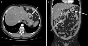
 Splenosis is acquired ectopic autotransplantation of viable splenic tissue, usually after traumatic splenic rupture or splenectomy.
The implants most often lie in the peritoneal or pelvic cavity but may occur in the liver, thorax, or other sites.
Clinical features are usually asymptomatic and incidental.
May rarely cause abdominal or pelvic pain, hemorrhage, bowel or urinary obstruction, torsion, or recurrent hematologic disease.
It can mimic metastatic disease, lymphoma, endometriosis, renal or hepatic tumors, and peritoneal carcinomatosis.
Diagnosis
A history of splenic trauma or splenectomy is the key clue.
On computed tomography (CT), magnetic resonance imaging (MRI), or ultrasonography, implants generally resemble the patient’s native splenic tissue in enhancement and signal characteristics, although imaging can remain indeterminate.
Preferred noninvasive tests are:
Technetium-99m heat-damaged red blood cell scintigraphy, often combined with single-photon emission computed tomography/CT.
Technetium-99m sulfur-colloid scintigraphy.
Spleen-specific MRI techniques, including ferumoxytol or superparamagnetic iron oxide–enhanced MRI in selected settings.
Management
Asymptomatic, confidently diagnosed splenosis generally requires no treatment.
Surgical excision is reserved for complications, significant symptoms, hypersplenism, or persistent diagnostic uncertainty after appropriate imaging.
Clinical takeaway: In a patient with prior splenic trauma or splenectomy and new abdominal, pelvic, hepatic, or pleural nodules, splenosis should be considered before biopsy or oncologic surgery; spleen-specific nuclear imaging is the usual next diagnostic step.