The spinothalamic tract is a sensory pathway to the thalamus.
It is a neural pathway that transmits sensory information about pain, temperature, and crude touch from the body to the brain.
It is part of the somatosensory system, which allows us to perceive and understand sensations from our environment.
The spinothalamic tract consists of two main pathways: the lateral spinothalamic tract and the anterior spinothalamic tract.
The lateral pathway carries information related to pain and temperature sensations, while the anterior pathway carries information related to crude touch and pressure sensations.
These pathways start in the spinal cord and project to the thalamus, which then relays the information to the appropriate regions of the brain for processing.
The spinothalamic tract consists of two pathways: anterior and lateral.
The anterior spinothalamic tract carries information about crude touch and firm pressure.
The lateral spinothalamic tract conveys pain and temperature.
The pathway decussates at the level of the spinal cord.
It is one of the three tracts which make up the anterolateral system.
There are two main parts of the spinothalamic tract:
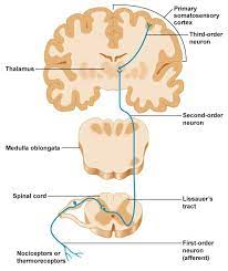
The spinothalamic tract uses three neurons to convey sensory information from the periphery to conscious level at the cerebral cortex.
Pseudounipolar neurons in the dorsal root ganglion have axons that lead from the skin into the dorsal spinal cord where they ascend or descend one or two vertebral levels via Lissauer’s tract and then synapse with secondary neurons in either the substantia gelatinosa of Rolando or the nucleus proprius.
Secondary neurons are called tract cells, whose axons cross over, or decussate, to the other side of the spinal cord via the anterior white commissure, and to the anterolateral corner of the spinal cord.
Decussation usually occurs 1-2 spinal nerve segments above the point of entry.
The axons travel up the length of the spinal cord into the brainstem, specifically the rostral ventromedial medulla.
The neurons ultimately synapse with third-order neurons in several nuclei of the thalamus—including the medial dorsal, ventral posterior lateral, and ventral posterior medial nuclei.
From there, signals go to the cingulate cortex, the primary somatosensory cortex, and insular cortex, respectively.
The ventral spinothalamic fasciculus is situated in the marginal part of the anterior funiculus and intermingled with the vestibulo-spinal fasciculus, and is derived from cells in the posterior column or intermediate gray matter of the opposite side.
Aβ fibers carry sensory information pertaining to crude touch from the skin.
After entering the spinal cord the first order neurons synapse, and the second order neurons decussate via the anterior white commissure.
These second order neurons ascend synapsing in the ventral posterolateral nucleus of the thalamus.
Incoming first order neurons can ascend or descend via the Lissauer tract.
The anterior spinothalamic tract fibers conduct information about pressure and crude touch data.
The fine touch is transmitted by fibers of the medial lemniscus.
The medial lemniscus is formed by the axons of the neurons of the gracilis and cuneatus nuclei of the medulla oblongata.
The medial lemniscus receives information about light touch, vibration and proprioception from the gracilis and cuneatus fasciculus of the spinal cord.
The cuneatus fasciculus receives the axons of the first order neuron which is located in the dorsal root ganglion that receives afferent fibers from receptors in the skin, muscles and joints.
The lateral spinothalamic tract/lateral spinothalamic fasciculus, which is a part of the anterolateral system, is an bundle afferent nerve bundle of fibers ascending through the white matter of the spinal cord, carrying sensory information to the brain.
The lateral spinothalamic tract carries pain, and temperature sensory information to the thalamus.
The lateral spinothalamic tract is composed primarily of fast-conducting, sparsely myelinated A delta fibers and slow-conducting, unmyelinated C fibers.
With the anterior spinothalamic tract, the lateral spinothalamic tract is sometimes termed the secondary sensory fasciculus or spinal lemniscus.
The neurons of the lateral spinothalamic tract originate in the spinal dorsal root ganglia, and project peripheral processes to the tissues in the form of free nerve endings which are sensitive to molecules indicative of cell damage.
The central processes enter the spinal cord in an area at the back of the posterior horn, the posterolateral tract.
The processes ascend approximately two levels before synapsing on second-order neurons.
Secondary neurons are situated in the posterior horn, specifically in the laminae regions I, IV, V and VI.
Region II is primarily composed of Golgi II interneurons, which are primarily for the modulation of pain, and largely project to secondary neurons in regions I and V.
Secondary neurons from regions I and V decussate across the anterior white commissure and ascend in the lateral spinothalamic tract.
These secondary neurons that decussate, ascend through the brain stem, including the medulla oblongata, pons and midbrain, as the spinal lemniscus until synapsing in the ventral posterior lateral nucleus of the thalamus.
The third order neurons from the thalamus project through the internal capsule and corona radiata to various regions of the cortex, primarily the main somatosensory cortex, Brodmann areas 3, 1, and 2.
Types of sensory information are accompanied by a compulsion to act: itch-scratch, pain-withdraw.
There are two sub-systems identified:
Direct, related to the direct conscious appreciation of pain.
Indirect, related to the affective and arousal impact of pain.
The anterolateral system is an ascending pathway that conveys pain,temperature and crude touch from the periphery to the brain.
It comprises three main pathways:
Spinothalamic tracts lateral and anterior, are important in the localization of painful or thermal stimuli.
The spinoreticular tract reticular formation causes alertness and arousal in response to painful stimuli.
The spinotectal tract orients the eyes and head towards the stimuli.
Second-order neuron axons in the spinothalamic tracts cross at every segmental level in the spinal cord, which aids in determining whether a lesion is in the brain or the spinal cord.
With lesions in the brain stem or higher, deficits of pain perception, touch sensation, and proprioception are all contralateral to the lesion.
With spinal cord lesions, however, the deficit in pain perception is contralateral to the lesion, whereas the other deficits are ipsilateral.
Unilateral lesions usually cause contralateral loss of pain and temperature.
Anaesthesia will normally begin 1-2 segments below the level of lesion, due to the sensory fibers being carried by dorsal-lateral tract of Lissauer up several levels upon entry into the spinal cord, and will affect all caudal body areas.
The spinothalamic tract (STT) is a sensory tract that carries nociceptive, temperature, crude touch, and pressure from our skin to the somatosensory area of the thalamus.
The spinothalamic tract (STT) is responsible for our quick withdraw reaction to a painful stimulus such as touching the stove burner.
The spinothalamic tract is composed of two adjacent pathways: anterior and lateral.
The anterior spinothalamic tract carries sensory input about crude touch.
The lateral spinothalamic tract carries information about pain and temperature.
The anterior and posterior spinothalamic tracts can be considered one pathway.
Three types of sensory fibers are associated with the spinothalamic tract: type III fibers, unmyelinated c-fibers, and myelinated A-delta fibers.
Peripheral receptors having associations with the spinothalamic tract pathway are nociceptors, thermal receptors, and thermal nociceptors.
Nociceptors are associated with A-delta and type III fibers, which are small, lightly myelinated axons for the transmission of fast, sharp pain.
Thermal receptors and thermal nociceptors are associated with A-delta and C fibers, which are small, unmyelinated axons that
conducti the transmission of slow-burning pain.
The spinothalamic tract pathway to the cerebral cortex starts with the dorsal root ganglions, which are composed of pseudounipolar neurons with the distal and proximal axonal processes.
Dorsal root ganglia lie adjacent to the spinal cord and are the first-order neuron of the spinothalamic tract pathway.
The first-order neurons enter the spinal cord through the lateral dorsal root entry zone to enter the Lissauer tract and synapses with second-order neurons in the substantia gelatinosa, located in the grey matter of the spinal cord.
The spinothalamic tract ascends in the ventrolateral aspect of the spinal white matter over the length of the spinal cord.
The spinothalamic tract of the anterolateral system terminates in the ventral posterolateral nucleus of the thalamus, the third-order neurons of this pathway.
From the thalamus, axons of VPL neurons project out of the thalamus laterally and course somatotopically through the internal capsule’s posterior limb of the and terminate in the postcentral gyrus primary somatosensory.
The main function of the spinothalamic tract is to carry pain and temperature via the lateral part of the pathway and crude touch via the anterior part.
The spinothalamic tract pathway is an essential sensory pathway for survival because it enables one to move away from noxious stimuli by carrying pain and temperature information from the skin to the thalamus where it is processed and transmitted to the primary sensory cortex.
The primary sensory cortex communicates with the primary motor cortex, to generate rapid movement in response to potentially harmful stimuli.
The spinothalamic tract has a role in responding to pruritogens, causing us to itch.
Itching suppresses the spinothalamic tract neuron response to the histamine effect.
The spinothalamic tract is important with any kind of spinal cord injury.
The spinothalamic tract is an anterolateral pathway,and is on the same side of the body.
A spinal lesion on this side of the body will cause a deficit of anything controlled below that point.
Several spinal cord syndromes have spinothalamic tract involvement.
Spinothalamic tract deficit leads to loss of pain and temperature sensations on one side of the body at about two levels below the lesion but on the contralateral side.
Te spinal cord syndrome Brown-Sequard syndrome, also known as a hemicord syndrome, compromises multiple tracts of the spinal cord, including the spinothalamic tract.
Bilateral loss of pain and temperature in a dermatomal fashion can be present in syringomyelia where the anterior white commissure becomes obliterated due to cavitation in the central spinal cord.
Similar clinical presentations to syringomyelia also present in acute central cervical cord syndrome.
The spinothalamic tract can be compromised due to trauma, cavitation and vascular issues.
The anterior spinal artery syndrome caused by infarction of the anterior spinal artery territory presents with pain and temperature loss bilaterally below the level of the lesion.
The vibratory and proprioceptive senses are preserved due to posterior column sparing.
Lateral medullary syndrome caused by occlusion of posterior inferior cerebellar artery affects the spinothalamic tract as it runs in the lateral aspect of the medulla: it results in loss of pain and temperature sensations over the entire contralateral side of the body as well as on the ipsilateral face due to the spinal trigeminal tract involvement.
The lateral pontine syndrome caused by the anterior inferior cerebellar artery can impact the spinothalamic tract giving a clinical picture that resembles that of lateral medullary syndrome.
In lateral medullary and pontine syndrome, cranial nerve deficits and cerebellar findings coexist with STT deficit findings as well as Horner syndrome.
A lesion localized to a ventral posterolateral in the thalamus will lead to an initial loss of all sensations from the contralateral side of the body, including pain.
Subsequently, the patient may experience pain in all, or part, of the contralateral side of the body; this is referred to as thalamic pain syndrome.
One reply on “Spinothalamic tract”
I’m amazed, I must say. Seldom do I come across a blog that’s
bot equally educative and engaging, and without a
doubt, you’ve hit tthe nail on the head. The issue is aan issue that too feew folks are speaking inteligently about.
Now i’m very happy I found this during my search for something concerning this. https://WWW.Waste-Ndc.pro/community/profile/tressa79906983/