 1113
1113
2724
The skull is a bony structure that forms the head.
It supports the structures of the face and provides a protective cavity for the brain.
The skull is composed of two parts: the cranium and the mandible.
The skull consists of two parts, of different embryological origin, the neurocranium and the facial skeleton.
The skull forms the anterior most portion of the skeleton.
Skull functions include:
protection of the brain,
fixing the distance between the eyes to allow stereoscopic vision
fixing the position of the ears to enable sound localization of the direction and distance of sounds.
It is made up of a number of fused flat bones, and contains many foramina, fossae, processes, and several cavities or sinuses.
The neurocranium, or braincase, is the protective cranial cavity that houses the brain and brainstem.
The upper areas of the cranial bones form the calvaria or skullcap.
The membranous viscerocranium includes the mandible.
The facial skeleton is formed by the bones supporting the face.
All of the bones of the skull are joined together, except the mandible, by suture.
The synarthrodial joints formed by bony ossification are immovable with Sharpey’s fibers permitting some flexibility.
There can be extra bone pieces within the suture known as wormian bones or sutural bones, and is found most commmonly in the course of the lambdoid suture.
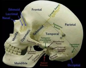
The human skull is generally considered to consist of twenty-two bones-eight cranial bones and fourteen facial skeleton bones.
The neurocranium is composed of the occipital bone, two temporal bones, two parietal bones, the sphenoid, ethmoid and frontal bones.
The bones of the facial skeleton are the vomer, two inferior nasal conchae, two nasal bones, two maxilla, the mandible, two palatine bones, two zygomatic bones, and two lacrimal bones.
The skull also contains, air-filled cavities known as paranasal sinuses, and numerous foramina.
The sinuses are lined with respiratory epithelium, reduce the weight of the skull, aid in the resonance of the voice and warm and moisturize the air drawn into the nasal cavity.
Skull openings are referred to as foramina.
The largest foramina of the skull is the foramen magnumit allows the passage of the spinal cord as well as nerves and blood vessels.
Skull processes of the skull include the mastoid process and the zygomatic processes.
The bones of the skull are formed both by intramembranous and endochondral ossification.
The skull roof bones, comprising the bones of the facial skeleton and the sides and roof of the neurocranium, are dermal bones formed by intramembranous ossification though the temporal bones are formed by endochondral ossification.
The endocranium, the bones supporting the brain, that is the occipital, sphenoid, and ethmoid bones are largely formed by endochondral ossification.
Thus frontal and parietal bones are purely membranous.
The anterior cranial fossa changes especially during the first trimester of pregnancy and skull defects can often develop during this time.
The skull is made up of 44 separate bony elements at birth.
During development the skull many of these bony elements gradually fuse together into solid bone.
Initially the bones of the skull are separated by dense connective tissue called fontanelles
There are six fontanelles: one frontal), one occipital), two anterolateral (sphenoid), and two posterolateral (mastoid).
The fontanelle regions are fibrous and moveable, necessary for birth and later growth of the skull.
As growth and ossification of the skull progresses, the connective tissue of the fontanelles is invaded and replaced by bone creating sutures.
The posterior fontanelle usually closes by eight weeks, but the anterior fontanel can remain open up to eighteen months.
The soft spot on a baby’s forehead, is the anterior fontanelle, located at the junction of the frontal and parietal bones
The baby’s pulse can be noted through the anterior fontanelle.
The skull in the neonate is large in proportion to other parts of the body.
The facial skeleton is one seventh of the size of the calvaria, but
in the adult it is half the size.
Craniosynostosis is a condition in which
If one or more of the fibrous sutures in an infant skull prematurely fuses it can cause craniosynostosis, changing the growth pattern of the skull.
With suture fusion the skull cannot expand perpendicular to the fused suture, it grows more in the parallel direction, and can result in an abnormal head shape and abnormal facial features.
With suture fusion craniosynostosis results in increased intracranial pressure leading possibly to visual impairment, sleeping impairment, eating difficulties, or an impairment of mental development.
With intense intracranial pressure there is disfigurement the internal surface of the skull, resulting in a copper beaten skull.
The copper beaten skull is most common in children.
The skull protects the brain from damage.
It is one of the least deformable structures found in nature with it needing the force of about 1 ton to reduce the diameter of the skull by 1 cm.
In some cases, the skull inability to deform following head injury can raise intracranial pressure through mechanisms such as a subdural hematoma.
When raised intracranial pressure occurs it can cause herniation of the brain out of the foramen magnum because there is no space for the brain to expand.
In adulthood male skulls tend to be larger and more robust than female skulls, which are lighter and smaller, with a cranial capacity about 10 percent less than that of the male.
Women’s skulls are slightly thicker and thus men may be more susceptible to head injury than women.
Male skulls tend to have more prominent supraorbital ridges, a more prominent glabella, more prominent temporal lines, larger, broader palates, squarer orbits, larger mastoid processes, larger sinuses, larger occipital condyles than those of females.
Male mandibles typically have squarer chins and thicker, rougher muscle attachments than female mandibles.
Female skulls generally have rounder orbits, and narrower jaws.
The cephalic index is the ratio of the width of the head, multiplied by 100 and divided by its length (front to back):
Dolichocephalic-long-headed
Mesaticephalic-medium-headed
Brachycephalic -short-headed