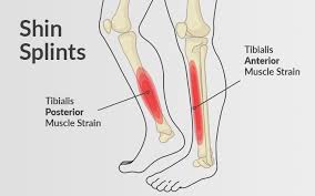
 A shin splint, (medial tibial stress syndrome), is pain along the inside edge of the shinbone, tibia, due to inflammation of tissue in the area.
It generally occurs between the middle of the lower leg and the ankle.
The pain may be dull or sharp.
Shin splint pain is described as a recurring dull ache, but sometimes is intense.
The pain increases during exercise, and some experience swelling in the pain area.
It is usually brought on by high-impact exercise that overloads the tibia.
It resolves during periods of rest.
Complications may include stress fractures.
Risk factors for shin splints:
Runners, dancers, gymnasts, military personnel.
Flat feet or rigid arches
Being overweight
Excessively tight calf muscles which can cause excessive pronation
Engaging the anti-pronatory muscles in excessive amounts of eccentric muscle activity.
Undertaking high-impact exercises on hard, non-compliant surfaces
People who have previously had shin splints are more likely to have them again.
Differential diagnosis:
Stress fracture, tendinitis, exertional compartment syndrome.
Treatment:
Rest with gradual return to exercise.
Other measures such as nonsteroidal anti-inflammatory drugs (NSAIDs), cold packs, physical therapy, and compression.
Shoe insoles may help some people.
Surgery is rarely required, but may be done if other measures are not effective.
Frequency at 4% to 35% at-risk groups.
The condition occurs more often in women.
Typically occur due to excessive physical activity.
Groups that are commonly affected include runners, dancers, gymnasts, and military personnel.
Diagnosis is based on the symptoms, with medical imaging done to rule out other possible causes.
Women are several times more likely to progress to stress fractures from shin splints than men, due to women having a higher incidence of diminished bone density and osteoporosis.
Shin splints has been attributed to the overloading of the lower leg, resulting in an increase in stress exerted on the tibia.
Such a sudden increase in intensity or frequency in activity level, fatigues muscles that help shock absorption, forcing the tibia to absorb most of the impact.
The lack of cushioning footwear, especially on hard surfaces, does not absorb transmitting forces while running or jumping.
It is this stress that is associated with shin splints.
The possibility of shin splints is also associated with muscle imbalance, weak core muscles, inflexibility and tightness of lower leg muscles, including the gastrocnemius, soleus, and planter muscles
The pain from shin splints is caused from a disruption of Sharpey’s fibers that connect the medial soleus fascia through the periosteum of the tibia where it inserts into the bone.
Repetitive impact forces stress and fatigue the soleus muscle and create tibial bending or bowing, contributing to shin splints.
Stress impact is worsened by running uphill, downhill, on uneven terrain, or on hard surfaces, improper footwear, including worn-out shoes, can also contribute to shin splints.
Diagnosis:
Shin splints are generally diagnosed from a history and physical examination.
Pressure paced over the tibia will recreate the type of pain experienced.
Magnetic resonance images of the lower leg show high signal areas around the tibia as signs of shin splints, with usually more than a 5 cm length of tibia is involved.
Clinical findings of swelling, redness, or poor pulses suggest a different diagnosis.
Differentiatial diagnosis:
stress fractures, compartment syndrome, nerve entrapment, and popliteal artery entrapment syndrome.
Bone scans and MRI can differentiate between stress fractures and shin splints.
Treatment:
Rest, ice, and gradually returning to activity.
Strengthening exercises should be performed after pain has subsided, on calves, quadriceps and gluteals.
Return to activity should be gradual, beginning with a short and low intensity level.
Running on other surfaces besides asphalt, such as grass, to decrease the amount of force the lower leg must absorb are encouraged.
Orthoses and insoles offset biomechanical irregularities, like pronation, and help to support the arch of the foot.
Other interventions include: exercise, footwear refitting, orthotics, manual therapy, balance training, cortisone injections, calcium and vitamin D supplementation, and deep tissue massage.
Less-common treatments for more-severe cases of shin splints include extracorporeal shockwave therapy (ESWT) and surgery.