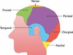
 The scalp is the anatomical area bordered by the human face at the front, and by the neck at the sides and back.
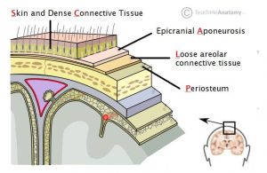
The scalp is usually described as having five layers, which can conveniently be remembered as a mnemonic:SCALP.
S: The skin on the head from which head hair grows, containing numerous sebaceous glands and hair follicles.
C: Connective tissue.-A dense subcutaneous layer of fat and fibrous tissue that lies beneath the skin, containing the nerves and vessels of the scalp.
A: The aponeurosis called epicranial aponeurosis, or galea aponeurotica, a tough layer of dense fibrous tissue which runs from the frontalis muscle anteriorly to the occipitalis posteriorly.
L: The loose areolar connective tissue layer provides an easy plane of separation between the upper three layers and the pericranium.
This connective tissue layer is torn off through this layer in the scalping process.
It also provides a plane of access in craniofacial surgery and neurosurgery.
Infectious agents can spread through it to emissary veins which then drain into the cranium.
The loose areolar tissue layer is made up of random collagen I bundles, collagen III, and is rich in glycosaminoglycans, allowing the more superficial layers of the scalp to shift about in relation to the pericranium.
The loose connective tissue area is hazardous because pus and blood spread easily within it, and can pass into the cranial cavity along the emissary veins.
Therefore, infections can spread from the scalp to the meninges, which could lead to meningitis.
P: The pericranium is the periosteum of the skull bones.
The pericranium provides nutrition to the bone and the capacity for repair.
The pericranium is lifted from the bone to allow a craniotomy.
The clinically important layer is the aponeurosis, as scalp lacerations through this layer means the anchoring of the superficial layers is lost and gaping of the wound occurs which would require suturing.
The blood supply of the scalp is via five pairs of arteries, three from the external carotid and two from the internal carotid:
Internal carotid:
the supratrochlear artery to the midline forehead, is a branch of the ophthalmic branch of the internal carotid artery.
the supraorbital artery to the lateral forehead and scalp as far up as the vertex, is a branch of the ophthalmic branch of the internal carotid artery.
external carotid
the superficial temporal artery gives off frontal and parietal branches to supply much of the scalp
the occipital artery which runs posteriorly to supply much of the posterior aspect of the scalp
the posterior auricular artery, a branch of the external carotid artery, ascends behind the auricle to supply the scalp above and behind the auricle.
The walls of the blood vessels in the fibrous tissue of the superficial fascial layer are firmly attached so cut ends of vessels do not readily retract, and even a small scalp wound may bleed profusely.
The veins of the scalp accompany the arteries and thus have similar names.
The scalp is innervated by the following nerves:
Supratrochlear nerve and the supraorbital nerve from the ophthalmic division of the trigeminal nerve
Greater occipital nerve (C2) posteriorly up to the vertex
Lesser occipital nerve (C2) behind the ear
Zygomaticotemporal nerve from the maxillary division of the trigeminal nerve supplying the hairless temple
Auriculotemporal nerve from the mandibular division of the trigeminal nerve
Lymphatic channels from the posterior half of the scalp drain to occipital and posterior auricular nodes.
Lymphatic channels from the anterior half drain to the parotid nodes.
The lymph eventually reaches the submandibular and deep cervical nodes.
Hair transplantation use techniques for the patient’s existing hair.
Candidates for this type of surgery are individuals who still have healthy hair on the sides and the back of the head for the transplant may be harvested from those areas.
The most utilized technique is the one known as micrografting.
The scalp is a common site for the development of lesions including:
Actinic keratosis
Basal-cell carcinoma
Epidermoid cyst
Merkel-cell carcinoma
Pilar cyst
Squamous cell carcinoma
Conditions associated with the scalp:
Cutis verticis gyrata
Dandruff
Head lice
Seborrhoeic dermatitis
Cradle cap, a form of seborrhoeic dermatitis which occurs in newborns
Tycoon’s cap, also known as acne necrotica miliaris, characterized by pustules and itching
The scalp has an important role in the aesthetics of the face.
Androgenic alopecia is male pattern hair loss from the scalp.
If the scalp is heavy and loose, a change with ageing, the forehead may be low, heavy and deeply lined.