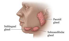
Within the mouth and throat 3 sets of salivary glands: the parotids, submandibular and sublingual glands plus multiple minor salivary glands.
Begin to form at 6-9 weeks gestation and the major salivary glands arise from ectodermal tissue.
There are three pairs of main salivary glands and between 800 and 1,000 minor salivary glands, all of which mainly serve the digestive process.
There are two types of salivary glands:
Serous glands: These glands produce a secretion rich in water, electrolytes, and enzymes (partied gland).
Mixed glands: These glands have both serous cells and mucous cells, and include sublingual and submandibular glands.
The largest of these are the parotid glands—their secretion is mainly serous.
The next pair are underneath the jaw, the submandibular glands, these produce both serous fluid and mucus.
The serous fluid is produced by serous glands in these salivary glands which also produce lingual lipase.
They produce about 70% of the oral cavity saliva.
Their secretion is mucinous and high in viscosity.
The third pair are the sublingual glands located underneath the tongue and their secretion is mainly mucous with a small percentage of saliva.
The minor salivary glands arise from either ectodermal or endodermal tissue, depending on their location.
The parotid gland lymph nodes are trapped within the gland and are located primarily in the superficial portion of the gland.
Lymphatic metastases may manifest within the substance of the parotid gland.
Salivary glands are made up of acini and ducts.
The acini secrete mucus, serum, or both and drain into the intercalated duct, followed by the striated duct, and finally into the excretory duct.
Myoepithelial cells surround the acini and intercalated duct and aid in expelling secretory products into the ductal system.
Parotid gland acini contain predominately serous cells.
Submandibular gland acini contain both mucous and serous cells.,
Sublingual and minor salivary glands have predominately mucous acini.
The parotid gland is the largest of the salivary glands, and located in a compartment anterior to the ear and is surrounded by fascia that suspends the gland from the zygomatic arch.
The parotid compartment contains the parotid gland, nerves, blood vessels, and lymphatic vessels, along with the gland itself.
The compartment is divided into superficial, middle, and deep portions, but the space has no discrete anatomic divisions.
The superficial portion of the parotid comparttment contains the facial nerve, great auricular nerve, and auriculotemporal nerve.
The VII cranial nerve runs through the parotid gland.
The middle portion of the parotid compartment contains the superficial temporal vein, which unites with the internal maxillary vein to form the posterior facial vein.
The deep portion of the parotid compartment contains the external carotid artery, the internal maxillary artery, and the superficial temporal artery.
The submandibular glands are the second largest salivary glands.
The submandibular glands are encapsulated glands located anterior and inferior to the angle of the mandible in the submandibular triangle formed from the anterior and posterior bellies of the digastric muscle and the inferior border of the mandible.
The submandibular gland has a superficial portion located lateral to the mylohyoid and a deep portion located between the mylohyoid and the hyoglossus., and the anterior facial vein pass superficially to the gland.
The facial artery crosses the deep portion of the gland, and the Wharton duct drains the gland.
Arterial blood supply is from the lingual and facial arteries and the anterior facial vein provides venous drainage.
The lymphatic drainage is from the submandibular nodes which then drain into to the deep jugular chain.
Submandibular and submaxillary glands are located between the digastric muscles and the mandible.
The submandibular glands can be palpable.
The sublingual glands are the smallest of the major salivary glands. Unlike the parotid and are unencapsulated.
The glands lies deep to the mucosa of the mouth floor.
The sublingual glands have 8-20 small ducts, which penetrate the floor of mouth mucosa to enter the oral cavity laterally and posteriorly to the Wharton duct.
The sublingual glands arterial supply is from the lingual artery, and lymphatic drainage is to the submental and submandibular lymph nodes, then to the deep cervical lymph nodes.
Minor salivary glands, approximately 600-1000 in number, are located throughout the paranasal sinuses, nasal cavity, oral mucosa, hard palate, soft palate, pharynx, and larynx.
Each gland is a individual in nature with its own duct opening into the oral cavity.
Salivary glands produce 1-1.5 L of saliva per day.
About 45% of saliva is produced by the parotid gland, 45% by the submandibular glands, and 5% each by the sublingual and minor salivary glands.
Saliva is produced at a low basal rate throughout the day.
During meals saliva production increases 10-fold increase in flow during meals.
Saliva functions to maintain lubrication of the mucous membranes and to clear food, cellular debris, and bacteria from the oral cavity, assists in initial digestion of food, and is protective for the teeth and prevents dental caries and enamel breakdown.

