 The retroperitoneum is divided into 3 anatomic regions that are utilized to guide therapy and describe the location of the retroperitoneal hematomas.
Zone 1: The central retroperitoneum from the diaphragm superiorly to the bifurcation of the aorta inferiorly. It contains the inferior vena cava, originals of the major renal visceral vessels, duodenum, and pancreas.
Zone 2: Includes both lateral perinephric areas of the upper retroperitoneum from the renal vessels medially to the lateral reflection of the posterior parietal peritoneum of the abdomen (from the diaphragm superiorly to the level of aortic bifurcation inferiorly). Organs contained include adrenal glands, kidneys, renal vessels, ureter, and ascending and descending colon.
Zone 3: Is inferior to the aortic bifurcation and includes the right and left internal and external iliac arteries and veins, distal ureter, and distal sigmoid colon and rectum.
Retroperitoneal bleeding refers to an accumulation of blood in the retroperitoneal space.
Retroperitoneal hematoma is defined as bleeding into the retroperitoneal space.
Retroperitoneal hematomas are the result of blood loss due to the injury of parenchymal tissue or vascular structures within the retroperitoneal cavity.
Retroperitoneal hematoma describes a heterogeneous condition that may arise spontaneously, as a result of trauma or iatrogenically. It is associated with high morbidity and mortality.
Retroperitoneal bleeding clinical is often occult and under-recognized by clinicians and is a cause of significant morbidity and mortality.
It is often occult and under-recognized by clinicians and is a cause of significant morbidity and mortality.
Often patients do not manifest clinically apparent signs and symptoms until a substantial amount of blood loss has occurred.
It is not uncommon for patients to present in frank hemorrhagic shock due to an underlying retroperitoneal hematoma.
Signs and symptoms may include abdominal or upper leg pain, hematuria, and shock.
Retroperitoneal bleeding can be caused by major trauma or by non-traumatic mechanisms.
Retroperitoneal bleeding, also referred to as retroperitoneal hematoma.
RB is most often caused by major trauma, such as from a traffic collisions or a fall.
Less common non-traumatic causes including:
Use of anticoagulants
A ruptured aortic aneurysm.
A ruptured renal aneurysm.
Acute pancreatitis.
Malignancy.
Iatrogenic RB can occur accidentally during medical procedures: cannulating the femoral artery for cardiac catheterization or for interventional radiology, and the administration of a psoas compartment nerve block.
Diagnosis comes from recognition of the above symptoms, and the accumulation of blood in the retroperitoneal space also compresses the femoral nerve.
Compression of the femoral nerve may lead to weakness in extension of the knee, with reduced patellar reflex, and weakness in flexing the hip.
It is not uncommon for patients to present in hemorrhagic shock.
The retroperitoneal space lies directly posterior to the peritoneal cavity.
The traumatic retroperitoneal hematoma heading can be subdivided into penetrating versus blunt.
The nontraumatic retroperitoneal hematoma category can be further broken down into spontaneous and iatrogenic.
The diagnosis of retroperitoneal hematoma is reliant upon the use of computed tomography (CT) scanning, which is often useful to confirm the diagnosis as well as identify the underlying cause.
Treatment modalities include observation, interventional radiology coiling/embolization, and operative management for unstable patients.
RB is the result of blood loss due to the injury of parenchymal tissue or vascular structures within the retroperitoneal cavity.
Blunt trauma comprises the majority of retroperitoneal hematomas.
The blunt mechanism results in compressive and deceleration forces, which often lead to crushing and shearing injuries to tissues and vascular structures: perirenal hematomas, pancreatic injuries, pelvic retroperitoneal hematomas as a result of pelvic fractures and disruption of nearby vascular structures, and direct avulsion injuries to blood vessels within the retroperitoneum.
Penetrating trauma leads to retroperitoneal hematoma is commonly the result of gunshot wounds or stabbings.
Injuries associated with penetrating trauma depend on specific organs or vessels affected by the trajectory of the knife or missile.
Retroperitoneal hematomas that occur outside the setting of trauma are either spontaneous or iatrogenic in etiology.
Iatrogenic retroperitoneal hematomas are the result of percutaneous interventions (PCI) or endovascular procedures.
Retroperitoneal hematoma is a rare complication of such procedures, but associated morbidity and mortality are high when it does occur.
Iatrogenic retroperitoneal hematomas are the result of percutaneous interventions or endovascular procedures: rare complication of such procedures-2018 study citing an incidence of 0.06%.
Risk factors for retroperitoneal hematoma following PCI: arterial puncture above the level of the inguinal ligament, female sex, treatment with GPIIb/IIIa inhibitors, and treatment with warfarin.
Spontaneous retroperitoneal hematoma is a relatively rare with a high degree of morbidity and mortality.
Spontaneous retroperitoneal hematoma is more common in the elderly, patients receiving anticoagulation therapy, and those with underlying coagulopathy: Some studies show 15% of elderly patients were receiving no blood thinning regimen.
The causes of spontaneous retroperitoneal hematoma include rupture of parenchymal lesions such as angiomyolipomas, cysts, and renal carcinomas or underlying vascular malformations such as aneurysm or pseudoaneurysm of retroperitoneal vessels: superior gluteal artery, various lumbar arteries, renal artery, and pancreaticoduodenal artery.
Traumatic retroperitoneal hematomas are more commonly the result of blunt injuries (67-80%) versus penetrating (20 to 33%).
It is estimated that renal injury affects up to 10% of those suffering blunt abdominal
Often there are no clinically apparent signs and symptoms until a substantial amount of blood loss has occurred.
It is not uncommon for patients to present in frank hemorrhagic shock due to an underlying retroperitoneal hematoma.
The retroperitoneal space lies directly posterior to the peritoneal cavity.
Retroperitoneal hematoma can be classified as traumatic versus nontraumatic.
The traumatic retroperitoneal hematoma heading can be further subdivided into penetrating versus blunt.
The nontraumatic retroperitoneal hematoma category can be further broken down into spontaneous and iatrogenic.
The diagnosis requires a high degree of clinical suspicion, and almost all cases, it is reliant upon the use of computed tomography (CT) scanning.
TREATMENT:
includes observation, interventional radiology coiling/embolization, and operative management for unstable patients.
Severe pelvic fractures often cause excessive bleeding, and extremely high mortality rates: the mortality rates of those undergoing angioembolization for pelvic hemorrhage was 17.6%, compared to the group that received no embolization with a mortality rate of 32.6%.
Blunt injuries to other retroperitoneal structures such as the duodenum and pancreas are rare in incidence, 0.2% and 5%, respectively, but carry high mortality rates, each cited above 20%.
Non-traumatic Retroperitoneal Hematoma
Retroperitoneal hematoma related to cardiac catheterization has been decreasing in incidence over the years, related to the rise in radial artery access.
Such patients who do suffer retroperitoneal hematoma, however, are at significantly higher risk for mortality and adverse cardiac events.
Spontaneous retroperitoneal hematoma is a rare occurrence confined mainly to case reports.
A retrospective study that identified 100 spontaneous retroperitoneal hematoma patients identified a 6% mortality rate directly attributed to retroperitoneal hematoma.
Hemorrhage should be suspected as the primary cause of hypotension/shock in a trauma patient.
Identification of potential bleeding sources based upon history, mechanism of injury, and the physical exam are helpful to identify potential etiologies.
A retroperitoneal bleed is considered as a source for shock in the trauma patient that has no other clear source for blood loss.
Physical exam should be conducted in the evaluation of all patients.
Findings consistent with hypovolemia/anemia, tachycardia and hypotension/poor peripheral perfusion,a bdominal tenderness may or may not be present.
Flank ecchymosis or Grey-Turner Sign, has poor sensitivity, present in 6.5% of cases in one study, and 1% in another.
In patients who suffer retroperitoneal hematoma after undergoing PCI manifest with abdominal, flank, and back pain, inguinal tenderness and swelling following the femoral approach.
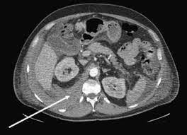
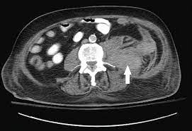
The mainstay of diagnosis for retroperitoneal hematoma is a contrast-enhanced CT-scan.
The hallmark feature of retroperitoneal hematomas, both traumatic and nontraumatic, is their occult nature.
The physical examination is commonly nondiagnostic.
CT scanning is part of a trauma workup, and will commonly reveal evidence of retroperitoneal hematoma, with sensitivities reportedly approaching 100%.
Sonography assessment in trauma exam is notoriously unreliable at detective presence of a retroperitoneal hematoma.
CT scan serves to identifies the presence, location, and to potentially therapy of retroperitoneal hematoma.
Additional evaluation of patients with retroperitoneal hematoma include: complete blood count, a metabolic panel to check for renal or electrolyte abnormalities, liver function tests to evaluate for hepatic dysfunction, coagulation studies, and a type and screen for transfusion requirements.
The management of retroperitoneal hematoma must be individualized, depending upon the etiology and overall clinical condition of each patient.
Treatment modalitie: include supportive, angioembolization/surgical exploration for more unstable patients.
Adequate resuscitation with blood products for acute hemorrhage should be undertaken.
Retroperitoneal injuries commonly occur in conjunction with intra-abdominal injuries.
With penetrating abdominal trauma, retroperitoneal hematoma develops as a result of direct injury to solid organs, viscera, or vasculature.
In the majority of these cases, surgical exploration is recommended to achieve hemostasis.
Persistence in hemodynamic instability, and the presence of an expanding or pulsatile hematoma, are indications fordefinitive surgical management.
The specific location of retroperitoneal hematoma in the setting of blunt injury is often able to be localized on contrast-enhanced CT scan, and managed based on findings.
The treatment of non-traumatic retroperitoneal hematoma is mainly based upon the location and characteristics of the bleeding.
Reversal of the coagulopathy should be undertaken to prevent further bleeding in patients with spontaneous retroperitoneal hematoma on anticoagulation.
Blood transfusion should be ordered for those with signs and symptoms or lab findings of anemia, and hemodynamic instability.
A majority of patients with spontaneous retroperitoneal hematoma and even those with post-procedural retroperitoneal hematoma do well with supportive care and blood transfusion alone.
In one series of retroperitoneal bleeding 24.7% of patients underwent an embolization procedure, and 6.7% underwent a surgical procedure, while 75% received a blood transfusion.
In another series of 100 patients with retroperitoneal hematoma, the rate of invasive management was only 16%.
The main predictor of the need for invasive therapy is contrast extravasation on CT scan.
Differential Diagnosis for traumatic retroperitoneal hemorrhage:
Acute abdomen
Perforated viscus
Solid organ injury
Vascular injury
Pelvic fracture
Abdominal compartment syndrome
Non-traumatic Retroperitoneal Hematoma
GI bleeding
Musculoskeletal pain
Appendicitis
Cholecystitis
Diverticulitis
Abdominal aortic aneurysm
Pancreatitis
Pyelonephritis
Prognosis
Traumatic retroperitoneal hematoma prognosis depends upon the extent and type of injury.
Blunt trauma-related retroperitoneal hematoma, mortality was 6.5% in the overall sample, with approximately 77% undergoing surgical management.
In a cohort dominated by penetrating trauma victims, mortality was noted to be 18%.
The retroperitoneal space lies directly posterior to the peritoneal cavity.
The retroperitoneal space into three different zones.
The central-medial zone (Zone I) falls between the two psoas muscles and contains midline structures such as the abdominal aorta, inferior vena cava, pancreas, and duodenum.
The perirenal zone (Zone II) begins lateral to the psoas muscles on either side and contains the kidneys, ureters, and portions of the colon.
The pelvic zone (Zone III) includes the bladder as well as a multitude of vascular structures, including a robust network for presacral veins.
Also, the retroperitoneum contains vital musculoskeletal structures such as the psoas muscles, vertebra, quadratus lumborum, and iliacus muscles. It houses connections to the diaphragm and bony pelvis.
Diagnosis classifies retroperitoneal hematoma as traumatic versus nontraumatic.
The traumatic retroperitoneal hematoma heading can be further subdivided into penetrating versus blunt.
The nontraumatic retroperitoneal hematoma category can be further broken down into spontaneous and iatrogenic.
The diagnosis of retroperitoneal hematoma requires a high degree of clinical suspicion.
Diagnosis is reliant upon the use of computed tomography (CT) scanning, which is often useful to confirm the diagnosis as well as identify the underlying cause.
Treatment modalities include observation, interventional radiology coiling/embolization, and operative management for unstable patients.
Etiology
Retroperitoneal hematomas are the result of blood loss due to the injury of parenchymal tissue or vascular structures within the retroperitoneal cavity.
In traumatic retroperitoneal hematoma, the mechanism of injury can be broken down into blunt or penetrating.
Blunt trauma comprises the majority of retroperitoneal hematomas seen in practice and, is the result of a transfer of energy from an outside source.
The blunt mechanism results in compressive and deceleration forces, which often lead to crushing and shearing injuries to tissues and vascular structures.
Penetrating trauma leading to retroperitoneal hematoma is commonly the result of lower energy mechanisms: gunshot wounds or stabbings.
Retroperitoneal hematomas that occur outside the setting of trauma.
Retroperitoneal hematoma, without trauma, are either spontaneous or iatrogenic in etiology.
Iatrogenic retroperitoneal hematomas are the result of percutaneous interventions or endovascular procedures: this is a rare complication of such procedures, associated morbidity and mortality are high when it does occur.
Risk factors for retroperitoneal hematoma following PCI appear to be arterial puncture above the level of the inguinal ligament, female sex, treatment with GPIIb/IIIa inhibitors, and treatment with warfarin.
Spontaneous retroperitoneal hematoma is a relatively rare clinical entity with a high degree of morbidity and mortality.
Spontaneous retroperitoneal hematoma suggest that this diagnosis is more common in the elderly, patients receiving anticoagulation therapy, and those with underlying coagulopathy.
While a majority of patients who suffer retroperitoneal hematomas are receiving anticoagulation therapy, however 15% are not.
Causes of spontaneous retroperitoneal hematoma: rupture of parenchymal lesions such as angiomyolipomas, cysts, and renal carcinomas or underlying vascular malformations such as aneurysm or pseudoaneurysm of any number of retroperitoneal vessels.
Traumatic retroperitoneal hematomas are more commonly the result of blunt injuries (67-80%) versus penetrating (20 to 33%).
The majority of these patients suffered from renal injuries as identified on computed tomography (CT) scan.
It is estimated that renal injury affects up to 10% of those suffering blunt abdominal trauma.
Large vascular injuries such as direct aortic injuries due to blunt trauma are rare.
Avulsion injuries to smaller aortic branches often occur and can be a cause of centromedial (Zone I) retroperitoneal hematoma.
Pelvic fractures comprise an estimated 2 to 8% of all fractures.
Severe fractures often cause excessive bleeding, and extremely high mortality rates.
Mortality rates of those undergoing angioembolization for pelvic hemorrhage were 17.6%, compared to the group that received no embolization with a mortality rate of 32.6%.
Blunt injuries to other retroperitoneal structures such as the duodenum and pancreas are rare in incidence (0.2% and 5%, respectively), but have high mortality rates, above 20%.
The etiology of non-traumatic retroperitoneal hematoma can be related to complications of percutaneous procedures or spontaneous in nature.
Retroperitoneal hematoma related to cardiac catheterization has been decreasing in incidence over the years, and is an infrequent complication.
Spontaneous retroperitoneal hematoma is a rare occurrence.
A retrospective study that identified 100 spontaneous retroperitoneal hematoma patients identified a 6% mortality rate directly attributed to retroperitoneal hematoma.
There must be a high degree of clinical suspicion for retroperitoneal hematoma, as there are no reliable features that are helpful in ruling in or out the diagnosis.
Most signs and symptoms of retroperitoneal hematoma are based upon the specific organs that are injured.
One should consider retroperitoneal bleeding as a source for shock in the trauma patient that has no other clear source for blood loss.
Presenting signs and symptoms of spontaneous retroperitoneal hematomas are often vague and nonspecific: abdominal pain seems to be the most common complaint (46-67.5%).
Other complaints include: pain in the back, flank, hip, and generalized weakness.
Symptoms of hypovolemia are common, such as syncope, pallor, and dizziness.
Physical exam findings consistent with hypovolemia/anemia are common in patients with ongoing blood loss from a retroperitoneal hematoma: tachycardia and hypotension/poor peripheral perfusion.
Abdominal tenderness may or may not be present with retroperitonel bleed.
Findings such as flank ecchymosis (Grey-Turner Sign) are of poor sensitivity, present in 6.5% of cases in one study, and 1% in another.
Retroperitoneal hematoma after undergoing PCI findings include abdominal, flank, and back pain.
Inguinal tenderness and swelling following the femoral approach are noted as being present in a majority of retroperitoneal hematoma cases.
Another common finding reported is that of femoral neuropathy, likely caused by nerve compression due to hematoma formation.
The mainstay of diagnosis for retroperitoneal hematoma is a contrast-enhanced CT-scan.
Retroperitoneal hematomas, both traumatic and nontraumatic, are occult in nature.
The physical examination is commonly nondiagnostic at best, and they are not readily detectable on plain film imaging or ultrasonography.
Patients who experience significant blunt and penetrating trauma will likely undergo extensive CT scanning as part of their trauma workup.
These scans will commonly reveal evidence of retroperitoneal hematoma, with sensitivities reportedly approaching 100%.
The bedside sonography in trauma is unreliable at detective presence of a retroperitoneal hematoma.
Evaluation of patients with no history of trauma a high degree of clinical suspicion is necessary to consider the diagnosis, and a CT scan is the diagnostic imaging modality of choice.
CT scan serves to identify presence, location, and to guide therapy of retroperitoneal hematoma.
The remainder of the evaluation of patients with retroperitoneal hematoma should include lab work commensurate with the patient’s overall presentation, complete blood count to assess for anemia, a metabolic panel to check for renal or electrolyte abnormalities, liver function tests to evaluate for hepatic dysfunction, coagulation studies to determine bleeding risk and a type and screen for transfusion requirements.
Treatment:includes supportive care with close observation for stable patients or angioembolization/surgical exploration for more unstable patients.
Resuscitation with blood products for acute hemorrhage may br required.
For patients presenting with penetrating abdominal trauma, retroperitoneal hematoma develops as a result of direct injury to solid organs, viscera, or vasculature, and In such cases, surgical exploration is recommended to achieve hemostasis.
The majority of patients with spontaneous retroperitoneal hematoma and even those with post-procedural retroperitoneal hematoma do well with supportive care and blood transfusion alone.
One of the greatest predictors of the need for invasive therapy was contrast extravasation on CT scan.
Differential Diagnosis
Traumatic Retroperitoneal Hematoma
Acute abdomen
Perforated viscus
Solid organ injury
Vascular injury
Pelvic fracture
Abdominal compartment syndrome
Non-traumatic Retroperitoneal Hematoma
GI bleeding
Musculoskeletal pain
Appendicitis
Cholecystitis
Diverticulitis
Abdominal aortic aneurysm
Pancreatitis
Pyelonephritis
Prognosis of traumatic retroperitoneal hemorrhage depends upon the extent and type of injury sustained.
Aortic injuries carry significantly higher morbidity and mortality than isolated kidney injuries causing a retroperitoneal hematoma.
In a cohort of primarily blunt trauma-related retroperitoneal hematoma, mortality was 6.5% in the overall sample, with approximately 77% undergoing surgical management.
In a cohort dominated by penetrating trauma victims, mortality was noted to be 18%.
Prognosis of spontaneous retroperitoneal hematoma is poor,
reflecting the fact that elderly patients with multiple comorbidities make up the majority of patients diagnosed with spontaneous retroperitoneal hematoma.
One retrospective study found a mortality of 10% at 30 days and 40% percent of those studied required ICU care, suggesting that nearly half of the cases are critical.
Complications of retroperitoneal hematoma include:
Infection/sepsis
Symptomatic anemia
Exsanguination
Abdominal compartment syndrome