 The pigmented layer of retina or retinal pigment epithelium (RPE) is the pigmented cell layer just outside the neurosensory retina.
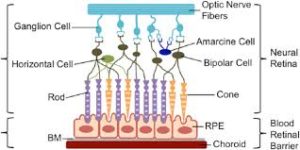
The retinal pigment epithelium (RPE) is a layer of cells located between the photoreceptor cells of the retina and the underlying choroid tissue.
The RPE nourishes retinal visual cells, and is firmly attached to the underlying choroid and overlying retinal visual cells.
The RPE is composed of a single layer of densely packed hexagonal cells with pigment granules.
The RPE functions include: light absorption, epithelial transport, spatial ion buffering, visual cycle, phagocytosis, secretion and immune modulation.
RPE are responsible for absorbing scattered light to improve the quality of the optical system, and light is concentrated by a lens onto the cells of the macula, resulting in a strong concentration of photo-oxidative energy.
Melanosomes absorb the scattered light and thus diminish the photo-oxidative stress.
The main function of the RPE is to support and nourish the photoreceptor cells, thus playing a crucial role in maintaining the health and function of the retina.
The RPE is heavily pigmented, containing melanin granules that help absorb excess light and prevent reflections within the eye.
This pigment also plays a role in protecting the retina from harmful ultraviolet light.
The RPE outer segments of photoreceptor cells undergo daily renewal, and acts as a phagocytic layer that engulfs and breaks down the old, discarded outer segments, recycling essential components for the photoreceptors and maintains their functionality.
It is involved in the transport of essential nutrients, ions, and molecules between the choroid and the photoreceptor cells.
The RPE serves as a metabolic bridge, ensuring a proper supply of oxygen, glucose, and other nutrients to support the energy-intensive work of the retina.
THE RPE also processes and removes waste products generated by the photoreceptor cells’ metabolism, maintaining the overall health of the retina by preventing the accumulation of toxic substances.
The RPE forms a barrier, known as the blood-retina barrier with tightly tightly packed cells that regulates the exchange of substances between the blood vessels in the choroid and the retina.
The blood-retina barrier barrier protects the delicate neural tissue of the retina from potentially harmful substances circulating in the bloodstream.
The RPE is involved in the processing and recycling of retinoids, which are derived from vitamin A, in the visual cycle.
Retinoids are crucial for the proper functioning of the visual system, especially for the regeneration of visual pigments in photoreceptor cells.
The high perfusion of retina brings a high oxygen tension environment, as the combination of light and oxygen brings oxidative stress.
RPE compose the outer blood–retinal barrier.
RPE epithelia has tight junctions between the lateral surfaces and implies an isolation of the inner retina from the systemic influences.
RPE tight junctions provides a barrier, a highly selective transport of substances for a tightly controlled environment.
RPE supply nutrients to photoreceptors, control ion homeostasis and eliminate water and metabolites.
In the eyes of albinos, the cells of this layer contain no pigment.
Dysfunction of the RPE is found in age-related macular degeneration, retinitis pigmentosa, and diabetic retinopathy.
Understanding the role of the retinal pigment epithelium is important for studying and treating various eye diseases, such as age-related macular degeneration, retinal dystrophies, and retinal detachments, which can significantly impact RPE function.
Gardner syndrome is characterized by FAP (familial adenomatous polyps), osseous and soft tissue tumors, retinal pigment epithelium hypertrophy and impacted teeth.