 A MUGA scan is a medical imaging test that is used to evaluate the function of the heart’s left ventricle.
MUGA stands for multiple-gated acquisition,and the test involves injecting a radioactive substance into a patient’s bloodstream prior to imaging.
As the radioactive material moves through the body, a special camera captures images of the heart’s left ventricle in motion.
These images are used to create a video that shows how the heart’s chambers fill and empty with each heartbeat, as well as how well the heart is pumping blood.
A MUGA scan can provide important information about cardiac function for a range of conditions, including coronary artery disease, heart valve problems, heart attack, and heart failure.
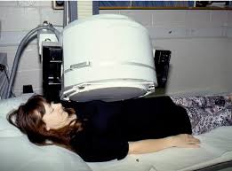
The test is noninvasive and generally takes about one hour to complete.
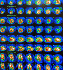
A MUGA scan involves an acquisition triggered or gated images at different points of the cardiac cycle.
This imaging provides a cine type of image of the beating heart, and allows the interpreter to determine the efficiency of the individual heart valves and chambers.
MUGA/Cine scanning is a adjunct to the echocardiogram.
The advantage of a MUGA scan over an echocardiogram or an angiogram is its accuracy.
An echocardiogram measures the shortening fraction of the ventricle and is limited by the user’s ability.
An angiogram is invasive and, often, more expensive.
A MUGA scan provides a more accurate representation of cardiac ejection fraction.
It is the preferred technique for measurement of left ventricular ejection fraction (LVEF) with a high degree of accuracy.
It an excellent correlation of MUGA-derived LVEF with values obtained by cardiac catheterization contrast ventriculography.
Radionuclide ventriculography is done to evaluate coronary artery disease (CAD), valvular heart disease, congenital heart diseases, cardiomyopathy, and other cardiac disorders.
MUGA is typically ordered for:
With known or suspected coronary artery disease, to diagnose the disease and predict outcomes
With lesions in their heart valves
With congestive heart failure
Who have undergone percutaneous transluminal coronary angioplasty, coronary artery bypass graft surgery, or medical therapy, to assess the efficacy of the treatment.
With low cardiac output after open-heart surgery
Who are undergoing cardiotoxic drug agents such as in chemotherapy with doxorubicin or immunotherapy, herceptin.
Who have had a cardiac transplant
Radionuclide ventriculography gives a much more precise measurement of left ventricular ejection fraction (LVEF) than a transthoracic echocardiogram (TTE).
Transthoracic echocardiogram is highly operator dependant, therefore radionuclide ventriculography is a more reproducible measurement of LVEF.
Its primary use today is in monitoring cardiac function in patients receiving certain chemotherapeutic agents (anthracyclines: doxorubicin or daunorubicin) which are cardiotoxic.
The MUGA scan is performed by labeling the patient’s red blood pool with a radioactive tracer, technetium-99m-pertechnetate (Tc-99m), and measuring radioactivity over the anterior chest as the radioactive blood flows through the large vessels and the heart chambers.
The introduction of the radioactive marker can either take place in vivo or in vitro.
In the in vivo method, stannous ions are injected into the patient’s bloodstream, and a subsequent intravenous injection of the radioactive substance, technetium-99m-pertechnetate, labels the red blood cells chnique is convenient, and more than 80 percent of the injected radionuclide usually binds to red blood cells with this approach.
In the in vitro method, some of the patient’s blood is drawn and the stannous ions are injected into the drawn blood, and technetium is subsequently added to the mixture.
Red blood cell binding of the radioactive tracer is generally more efficient than in vitro labeling, and it is preferred in patients with indwelling intravenous catheters to decrease the adherence of Tc-99m to the catheter wall and increase the efficiency of blood pool labeling.
A gamma camera detects the low-level 140 keV gamma radiation being given off by Technetium-99m (99mTc).
As the gamma camera images are acquired, the patient’s heart beat is used to ‘gate’ the acquisition, resulting is a series of images of the heart one at each stage of the cardiac cycle: usually 16 images.
The test can be performed as either a resting or a stress MUGA.
The stress MUGA measures the heart performance during exercise and is usually performed to assess the impact of a suspected coronary artery disease.
In some cases, a nitroglycerin MUGA may be performed, where a vasodilator is administered prior to the scan.
Volumetrically derived blood pools in the chambers of the heart and timed images may be computationally interpreted to calculate the ejection fraction and injection fraction of the heart, and can calculate ventricle volumes.
It is an accurate, inexpensive and easily reproducible means of measuring and monitoring the ejection and injection fractions of the ventricles, in assessing global heart performance.
While it exposes patients to less radiation than do comparable chest x-ray studies, the radioactive material is retained in the patient for several days after the test.
Radionuclide ventriculography has largely been replaced by echocardiography, which is less expensive, and does not require radiation exposure.
In normal subjects, the left ventricular ejection fraction (LVEF) should be about 50% (range, 50-80%).
There should be no area of abnormal wall motion: hypokinesis, akinesis or dyskinesis.
Abnormalities in cardiac function may be manifested as a decrease in LVEF and/or the presence of abnormalities in global and regional wall motion.
An uneven distribution of technetium in the heart indicates that the patient has coronary artery disease, a cardiomyopathy, or blood shunting within the heart.
Abnormalities in a resting MUGA usually indicate a heart attack, while those that occur during exercise usually indicate ischemia.
In a stress MUGA patients with coronary artery disease may exhibit a decrease in ejection fraction.
The MUGA scan can help pinpoint the site in the heart that has sustained damage as well as assess the degree of damage.
MUGA scans are also used to evaluate heart function prior to and while receiving certain chemotherapies (doxorubicin) or immunotherapy (Trastuzumab) that have a known effect on heart function.
The Massardo method estimates the volume of the ventricles and thus ultimately the ejection fraction.
The Massardo method enables a 3D volume to be estimated from a 2D image of decay counts.
The ejection fraction can then be calculated:
EDV/ESV-where the EDV (end-diastolic volume) is the volume of blood within the ventricle immediately before a contraction and the ESV (end-systolic volume) is the volume of blood remaining in the ventricle at the end of a contraction: The ejection fraction is hence the fraction of the end-diastolic volume that is ejected with each cycle.
Average sensitivity approximately 90% and specificity 76% for coronary artery disease.