Calcium pyrophosphate deposition disease (CPDP).
Calcium pyrophosphate dihydrate (CPPD) crystal deposition disease, also known as pseudogout and pyrophosphate arthropathy, is a rheumatologic disease which is thought to be secondary to abnormal accumulation of calcium pyrophosphate dihydrate crystals within joint soft tissues.
Affects 10-15% of individuals from 65-75 years of age.
Affects up to 60% of individuals 85 years and older.
Women are at a slightly higher risk than men to have calcium pyrophosphate dehydrate crystal deposition disease, with an estimated ratio of occurrence of 1.4:1.
Mean age of presentation is 70 years with women more likely than men to be effective.
If it occurs before age 50 years a metabolic disease: hyperparathyroidism, hemochromatosis, hypophosphatasia, and hypomagnesaemia must be considered.
Very few patients have associated metabolic abnormalities or heritable diseases.
Deposition of crystals lead to neutrophil phagocytosis and chemotactic recruitment of more inflammatory cells.
The symptoms can be monoarticular or polyarticular.
Symptoms usually last for days to weeks, and often recur.
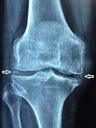
Although any joint may be affected, the knees, wrists, and hips are most common.
May be asymptomatic.
The knee joint is most commonly affected.
When symptomatic, the disease is similar to a gout attack:
severe pain
warmth
swelling of one or more joints
Many patients have acute or chronic arthritis resembling gout.
Up to two thirds of patients with CPDD disease have radiographic evidence of involvement of the cervical spine but are asymptomatic.
The findings are those of odontoid crown- like densities.
Involvement of the cervical spine is referred to as a crowned dens syndrome.
CPPD is treated with nonsteroidal anti-inflammatory drugs, colchicine or a short course of corticosteroids.
An acute inflammatory crystal induced arthritis may mimic infectious process such as septic arthritis.
Strongly associated with osteoarthritis and rheumatoid arthritis, as well as metabolic conditions such as hyper parathyroidism, gout,hemachromatosis, chronic kidney disease, and hypomagnesemia.
There can also be findings of osteoarthritis.
The cause of CPPD disease is unknown.
Increased breakdown of adenosine triphosphate resulting in increased pyrophosphate levels in joints, is thought to be one reason why crystals may develop.
Familial cases are rare.
A genetic study found an association between CPPD and a region of chromosome 8q.
The ANKH gene is involved in crystal-related inflammatory reactions and inorganic phosphate transport.
CPP disease is defined by presence of joint inflammation and the presence of CPPD crystals within the joint.
The crystals are can be detected by imaging and/or joint fluid analysis.
Medical imaging: x-ray, CT, MRI, or ultrasound may detect chondrocalcinosis within the affected joint, indicating a substantial amount of calcium crystal deposition within the cartilage or ligaments.
Ultrasound is also a reliable method to diagnose CPPD.
Using ultrasound, chondrocalcinosis may be depicted as echogenic foci with no acoustic shadow within the hyaline cartilage or fibrocartilage.
By x-ray, CPPD can appear similar to other diseases: ankylosing spondylitis and gout.
The white blood cell count is often raised with pseudogout.
Diagnosis requires the presence of synovial fluid CPP crystals and polarizing light microscopy with radiologic evidence of chondrocalcinosis as supporting evidence.
Arthrocentesis, allows the testing of the synovial fluid for the calcium pyrophosphate crystals that are present in CPPD.
When stained with H&E stain, calcium pyrophosphate crystals appears deeply blue.
CPP crystals have rhomboid shape and weak positive birefringence on polarized light microscopy.
Birefringence on polarized light microscopy is the most reliable method of identifying the crystals under the microscope.
Birefringence on polarized light microscopy suffers from poor sensitivity, specificity, and inter-operator agreement.
Imaging: X-ray, CT, or other imaging usually shows accumulation of calcium within the joint cartilage, known as chondrocalcinosis.
The sensitivity of routine laboratory testing for calcium pyrophosphate deposition disease ranges from 12 to 83%.
An acute CPPD arthrophy attack can have synovial fluid leukocyte concentration between 9000 and 60,000 cells per millimeter cubed.
Treatment is not advised if the condition is not causing pain: because of the risks of anti inflammatory agents.
For acute pseudogout, treatments include intra-articular corticosteroid injection, systemic corticosteroids, non-steroidal anti-inflammatory drugs (NSAIDs), or colchicine.
NSAIDs are administered in low doses to help prevent CPPD.
In refractory disease hydroxychloroquine or methotrexate may be beneficial.
The condition is more common in older adults.
It is estimated to affect 4% to 7% of the adult population.
While it may cause considerable pain, but it is never fatal.