 Heavy charged particles that can be deposited at specific tissue sites with precision.
Protons are extracted from proton donor materials by a synchrotron or cyclotron, and accelerated through a circular, evacuated conduit or cavity, using powerful magnets to shape their path, until they reach the energy required to just traverse a human body, usually about 200 MeV.
Compared with conventional x-ray treatment, low-dose radiation to normal tissue while the dose to tumors remains high, which can decrease treatment-related toxic effects and improve cancer outcomes.
The chief advantage of proton therapy over other types of external beam radiotherapy is that the dose of protons is deposited over a narrow range of depth; hence in minimal entry, exit, or scattered radiation dose to healthy nearby tissues.
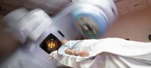
Proton therapy is a type of external beam radiotherapy that uses ionizing radiation from a particle accelerator to target a tumor with a beam of protons.
These charged particles damage the DNA of cells, ultimately killing them by stopping their reproduction and thus eliminating the tumor.
Cancer cells are vulnerable to attacks on DNA because of their high rate of division and their limited ability to repair DNA damage.
Allows ultra precise treatment for recurrent head and neck cancers, and with diseases that are near critical sites such associated the optic nerve or cavernous sinus.
Some cancers with specific defects in DNA repair may be more sensitive to proton radiation.
The process directs protons at the tumor target, where the bulk of the radiation dose is to a finite depth in tissue with minimal residual radiation beyond the target.
Like photon radiation therapy, proton therapy is often used in conjunction with surgery and/or chemotherapy to most effectively treat cancer.
The vast majority of the proton radiation is delivered to the tumor, not to the skin and shallow tissues in front of the tumor or to the deep tissues behind the tumor.
In some machines, which deliver protons of only a specific energy, a custom mask made of plastic is interposed between the beam source and the patient to adjust the beam energy to provide the appropriate degree of penetration.
Proton therapy allows delivery of a highly conformal beam, conforming to the shape and depth of the tumor and sparing much of the surrounding, normal tissue.
Proton therapy can give similar or higher radiation doses to the tumor with a 50%-60% lower total body radiation dose compared to traditional RT.
Protons can focus energy delivery to fit the tumor shape, delivering only low-dose radiation to surrounding tissue.
Proton dose delivered to tissue is maximized only over the last few millimeters of the particle’s range; this maximum is called the spread out Bragg peak.
Accelerators used for proton therapy typically produce protons with energies of 70 to 250 MeV.
Tissue closer to the surface of the body than the tumor gets less radiation, and thus less damage.
Tissues deeper in the body get very few protons, so the dose becomes immeasurably small.
Tissues behind, or deeper, than the tumor get almost no radiation, the tissues in front of the tumor get radiation dosage based on the Bragg peak.
The Bragg peak of ejected protons gives proton therapy advantages over other forms of radiation, since most of the proton’s energy is deposited within a limited distance.
This allows for conformal dose distributions to be created around even very irregularly shaped targets, and for higher doses to targets surrounded or backstopped by radiation-sensitive structures such as the optic chiasm or brainstem.
Has virtually no scatter.
Protons release a relatively small radiation dose when entering the body, and moves through tissues until out of energy.
Protons release their remaining ionizing radiation at the end of their energy range, the Bragg peak.
Has become a standard of care for several cancers, but it has not been proven that it has reduced toxic effects or increased survival, compared with conventional therapies.
Presently, is the preferred radiation modality for many pediatric, base of skull, and primary liver cancers.
A systematic review and metaanalysis showed a statistically improved five year overall survival for early stage NSCLC for proton beam radiotherapy compared with photon beam radiotherapy 60%, versus 41.3%, respectively: local failure, regional failure, and distant metastases were comparable.
Most proton therapy systems use isochronous cyclotrons.
Linear accelerators, as also used for photon radiation therapy.
Modern proton systems incorporate high-quality imaging for daily assessment of tumor contours, treatment planning software with 3D dose distributions, and various system configurations.
Pencil beam scanning, gives therapy by sweeping a proton beam laterally over the target so that it gives the required dose while closely conforming to shape of the targeted tumor.
Proton therapy is used to treat conditions in two broad categories:
Disease sites that respond well to higher doses of radiation.
Dose escalation has sometimes shown a higher probability of response than conventional radiotherapy.
These include, among others, uveal melanoma, skull base and paraspinal tumor (chondrosarcoma and chordoma), and unresectable sarcoma.
In all these cases proton therapy gives significant improvement in the probability of local control, over conventional radiotherapy.
For eye tumors, proton therapy also has high rates of maintaining the natural eye.
Irreversible long-term side effects of conventional radiation therapy for pediatric cancers: growth disorders, neurocognitive toxicity, ototoxicity,effects on learning and language development, and renal, endocrine and gonadal dysfunctions.
There is minimal exit dose when using proton radiation therapy, dose to surrounding normal tissues is significantly limited, reducing the acute toxicity, and risk for these long-term side effects.
With cancers requiring craniospinal irradiation, the absence of exit dose with proton therapy, the heart, mediastinum, bowel, bladder and other tissues anterior to the vertebrae is eliminated, reducing of acute thoracic, gastrointestinal and bladder side effects.
Proton therapy is an option for retinoblastoma and intraocular melanoma.
Proton therapy has been described as the primary treatment for ocular melanoma.
Momentum cooling technique in proton therapy for eye treatment can significantly enhance its effectiveness, and reduces the radiation dose administered to healthy organs while ensuring that the treatment is completed within a few seconds: it improves comfort during the procedure.
The side effects of the radiation to skull based tumors include: pituitary hormone dysfunction and visual field deficit, cranial neuropathy, radiation-induced osteosarcoma and osteoradionecrosis: these side effects are eliminated with proton therapy for skull base tumors.
For recurrent head and neck cancer requiring reirradiation, proton therapy is able to maximize a focused dose of radiation to the tumor while minimizing dose to surrounding tissues, hence a minimal acute toxicity profile, even in patients who got multiple prior courses of radiotherapy.
A study showed that proton therapy has low toxicity to nearby healthy tissues and similar rates of disease control compared with conventional radiation for breast cancer: proton pencil beam scanning techniques can reduce both the mean heart dose and the internal mammary node dose to essentially zero.
Advanced radiation therapy technologies such as proton therapy may offer significant and clinically relevant advantages such as sparing important organs at risk and decreasing the risk for late normal tissue damage while still achieving the primary goal of disease control in lymphoma.
This is especially important for lymphoma patients who are being treated with curative intent and have long life expectancy following therapy.
In prostate cancer cases, proton therapy has been found to have a reduction in long term rectal and genito-urinary damage when treated rather than photons, meaning X-ray or gamma ray therapy.
Others studies showed only small differences.
The relatively small improvement found may be the result of inconsistent patient set-up and internal organ movement during treatment, which offsets most of the advantage of increased precision.
Dose errors of around 20% can result from motion errors of just 2.5 mm and the prostate motion is between 5–10 mm (0.20–0.39 in).
Proton therapy has great potential to increase therapeutic tolerance for patients with GI malignancy: decreasing radiation dose to organs at risk may also help facilitate chemotherapy dose escalation or allow new chemotherapy combinations.
Hepatocellular carcinoma: proton therapy gives favorable results related to local tumor control, progression-free survival, and overall survival.
Reirradiation for recurrent cancer
Re-irradiation is a potentially curative treatment option for patients with locally recurrent head and neck cancer.
Proton therapy in the setting of concurrent chemoradiotherapy is associated with fewer 90-day unplanned hospitalizations and overall survival compared with concurrent photon therapy and chemoradiotherapy.
Proton beam therapy versus IMRT for locally advanced esophageal : proton beam therapy reduced the risk and severity of adverse events compared with IMRT while maintaining similar progression free survival.
Proton therapy is associated with a lower risk of a second cancer.
Proton therapy is a type of external beam radiotherapy, and shares risks and side effects of other forms of radiation therapy.
The dose outside of the treatment region can be significantly less for deep-tissue tumors than X-ray therapy, because proton therapy takes full advantage of the Bragg peak.
Proton therapy deposits the bulk of the energy at the last few millimeters of its range and proton cranial spinal irradiation results in minimal RT dose beyond the neuraxis and is associated with significantly fewer toxic effects compared with photon based craniospinal radiation.