Polymorphous light eruption (PLE), is a non-life-threatening and potentially distressing skin condition that is triggered by sunlight and artificial UV exposure.
About one-quarter of people being investigated for a photosensitivity disorder are diagnosed with PLE.
The prevalence in the general population is 10 to 15% and may even be as high as 40%.
It is also more prevalent in Central Europe and Scandinavia.
PLE is more common in young adults and has a female preponderance with a ratio of 2:1 female-to-male.
It occurs in a genetically susceptible person.
It is suggested that an undefined endogenous or exogenous photo-allergen may trigger a delayed immune reaction resulting in PLE.
Occurs particularly in temperate climates during the spring and early summer.
Its clinical appearances are polymorphic.
Its itch can be significant.
It lasts several days with annual recurrences.
It is an idiopathic primary photodermatosis.
Typically, the first episode develops in the spring following the first exposure to intense sunlight.
Subsequent episodes of the rash occur several hours to days following subsequent sun exposure.
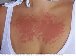
PLE appears on areas of the skin newly exposed to sunlight: neckline, backs of hands, arms and legs, and feet, but less commonly the face.
With PLE there may be feelings of burning and severe itching.
Clinically smooth red small papules that merge into plaques, small fluid-filled blisters and less commonly target-shaped lesions which look like erythema multiforme may be visible.
It may occur in other parts of the body in patients treated for inflammatory skin diseases with phototherapy.
The rash is usually symmetrical and characteristically similar with each recurrence.
Fever, fatigue and headaches associated with the eruption, are rare.
The rash may persist for many days to a couple of weeks.
The rash resolves spontaneously without scarring as long as further sunlight exposure is avoided.
The eruption can last longer than a few days if persistent and repeated sun exposure occurs.
The cause of PLE is not established.
It is thought to be due to a type IV delayed-type hypersensitivity to an allergen produced in the body following sunlight exposure.
It occurs in genetically susceptible persons.
It is also thought that skin microbiome could be involved in pathogenesis of the disease.
PLE can be provoked by UVA or UVB rays.
PMEcan be triggered even by sunlight through glass. UV-A is a major constituent of sunlight, can pass through glass, is relatively resistant to sunscreen and can cause light eruption without sunburn.[1]
Artificial UV light sources from tanning units and phototherapy treatment units can also trigger PLE.
About three-quarters of sufferers acquire PLE after UV-A exposure only.
One-tenth acquire PLE after UV-B exposure only, and the rest after a combination of UV-A and UV-B exposure.
People vary in the amount of sun exposure needed to trigger the rash.
Oxidative stress and the modification of the redox status of the skin has been implicated in the expression of PLE.
Half of individuals with PLE have a family history, demonstrating a clear genetic influence.
A natural fall in estrogens may account for the tendency for PLE to remit after the menopause.
Any investigations as to diagnosis are usually to exclude other conditions, particularly lupus and porphyria.
Photoprovocation tests are usually not required but may be undertaken by specialized centers: about small area of the frequently affected skin is exposed to varying doses of UVA and minimal erythema dose (MED) and of broadband UVB for three consecutive days.
An examination of the skin to detect the rash is made, however, up to 40% have false negative responses.
Biopsy findings: edema in the epidermis with a lymphocytic infiltrate without vasculitis, neutrophils, spongiosis and vesicle formation.
Similar findings occur in
solar urticaria, photosensitive dermatitis, and photosensitivity drug reaction.
Photosensitivity is also found in some of the porphyrias: nearly all cases of porphyria cutanea tarda exhibit blister formation on the skin within 2–4 days of light exposure.
Variegate porphyria and hereditary coproporphyria can also exhibit symptoms of light-induced blisters.
Variants of PLE:
Juvenile spring eruption is a cutaneous condition that affects the helices of the ears, and is seen particularly in boys.
Benign summer light eruption is a clinically short-lived, itchy, papular eruption particularly affecting young women after several hours of sunbathing at the beginning of summer or on sunny vacations.
Actinic Prurigo is a hereditary form of PLE occurring typically in native Americans.
TREATMENT/MANAGEMENT:
Steadily increasing exposure to sunlight, reduces light sensitivity with repeated sun exposure.
Covering up with clothing and applying a water-resistant sun protection factor (SPF) 50+ sunblock cream before sun exposure and then every two hours thereafter confers some protection, application of topical corticosteroids may lessen the redness and itch, and for preventing predictable flare-ups, short courses of oral corticosteroids are sometimes considered.
Other treatment options; low dose phototherapy, administration of hydroxychloroquine.
Generally, PLE resolves without treatment.
PLE lesions generally leave no scar.
There is a possible link with autoimmune thyroid disease, and some progress to a autoimmune disease.
In Germany the female to male ratio has been cited as 9:1.
It occurs in all age groups and all skin types.
Those experiencing sun exposure all year round seldom acquire PLE eruption.
It is less common near the equator.
This condition are most common between the spring and autumn months in the northern hemisphere and at higher altitudes.
Psychological distress is noted in more than 40% of peoples with PLE.