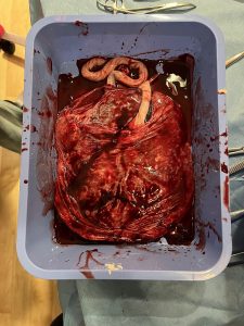
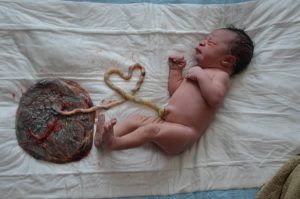
Placental expulsion, also called afterbirth, occurs when the placenta comes out of the birth canal after childbirth.
The period from just after the baby is expelled until just after the placenta is expelled is called the third stage of labor.
The third stage of labor can be managed actively with or it can be managed expectantly.
Managed expectantly, is also known as physiological management or passive management: allowing the placenta to be expelled without medical assistance.
In some cultures the placenta is kept and consumed by the mother over the weeks following the birth (placentophagy).
With fetal hypothalamus maturity activation of the HPA axis (hypothalamic-pituitary-adrenal axis) initiates labour through two hormonal mechanisms.
The mechanisms lead to contractions in the myometrium, a mechanical cause of placental separation, which is due to the shear force and contractile & involution changes that occur within the uterus distorting the placentome.
ACTH increases fetal cortisol, which acts by increasing Prostaglandin F2α, which both abolishes the progesterone block, and lowers the oxytocin receptor threshold; and increases expression of relaxin, stretching the pelvic ligaments
Increases expression of placental gene transfer in the fetal trophoblast cells.
Placental gene transfer produces prostaglandin E2, which is a catalyst for pregnenolone to C-19 steroids, such as estrogen.
Estrogen increases vaginal lubrication,
softens collagen fiber structures in the cervix, vaginal, and associated tissues, and increases contraction associated proteins-connexins.
As the HPA axis activates, the posterior pituitary of the fetus increases production of oxytocin, which stimulates the maternal myometrium to contract.
In the seventh month of pregnancy the MHC-I complexes increase in the interplacentomal arcade reduces the bi- and tri-nucleate cells, which are a source of immune suppression in pregnancy.
By the ninth month the endometrial lining has thinned, due to loss of trophoblast giant cells, which exposes the endometrium directly to the fetal trophoblast epithelium.
An increase in maternal MHC-I, T-helper 1 (Th1) cells, and macrophages induced apoptosis of trophoblast cells and endometrial epithelial cells, facilitating placental release.
Th1 cells attract an influx of phagocytic leukocytes into the placentome at separation, allowing further degration of the extracellular matrix.
After delivery, loss of fetal blood return to the placenta accounts for shrinkage and collapse of the villi with subsequent fetal membrane separation.
Active management includes umbilical cord clamping, stimulation of uterine contraction and cord traction.
Umbilical cord clamping involves clamping of the umbilical cord, often within seconds or minutes of birth.
Uterine contraction assists in delivering the placenta, reduces the placental surface area, often forming a temporary hematoma at their former interface.
Myometrial contractions can be induced with medication, usually oxytocin via intramuscular injection.
The use of ergometrine, on the other hand, is associated with nausea or vomiting and hypertension.
Breastfeeding after birth stimulates oxytocin which increases uterine tone, and through uterine massage also causes uterine contractions.
Controlled cord traction (CCT) consists of pulling on the umbilical cord while applying counter pressure to help deliver the placenta.[3] It may be uncomfortable for the mother. Its performance requires specific training. Premature cord traction can pull the placenta before it has naturally detached from the uterine wall, resulting in hemorrhage. Controlled cord traction requires the immediate clamping of the umbilical cord.
A Cochrane review came to the conclusion that controlled cord traction does not clearly reduce severe postpartum hemorrhage but overall resulted in a small reduction in postpartum hemorrhage and mean blood loss.
It did reduce the risk of manual placenta removal.
Controlled cord traction should be recommended if the care if the provider has the skills to administer it.
Manual placenta removal is usually carried out under anesthesia or more rarely, under sedation and analgesia.
A hand is inserted through the vagina and cervix into the uterine cavity and the placenta is detached from the uterine wall and then removed manually.
A placenta that does not separate easily from the uterine surface suggests the presence of placenta accreta.
The of postpartum bleeding will be reduced in women offered active management of the third stage of labour.
Cochrane study results indicate active management of the third stage of labour, consisting of controlled cord traction, early cord clamping plus drainage, and a prophylactic oxytocic agent, reduced postpartum hemorrhage by 500 or 1000 mL or greater, as well as related morbidities including mean blood loss, incidences of postpartum hemoglobin becoming less than 9 g/dL, blood transfusion, need for supplemental iron postpartum, and length of third stage of labour.
Active management increased adverse effects such as nausea, vomiting, and headache.
A retained placenta is one that does not undergo expulsion within a normal time limit.
Risks of retained placenta include: hemorrhage and infection.
If the placenta fails to deliver in 30 minutes in a hospital environment, manual extraction may be required if heavy ongoing bleeding occurs, and very rarely a curettage is necessary to ensure that no remnants of the placenta remain.
However, it is common for licensed care providers to wait for the placenta’s birth up to 2 hours in some instances.