 The pharynx is the part of the throat behind the mouth and nasal cavity, and above the esophagus and trachea.
The pharynx carries food and air to the esophagus and larynx respectively.
The pharynx is made up of three parts.
The lower two parts—the oropharynx and the laryngopharynx are involved in the digestive system.
The laryngopharynx connects to the esophagus and it serves as a passageway for both air and food.
Air enters the larynx anteriorly but anything swallowed has priority and the passage of air is temporarily blocked.
The flap of cartilage called the epiglottis stops food from entering the larynx.
The pharynx is part of the digestive system and the conducting zone of the respiratory system.
The conducting zone of the respiratory system includes the nostrils of the nose, the larynx, trachea, bronchi, and bronchioles.
The conducting zone of the respiratory system filters, warms and moistens air and conducts it into the lungs
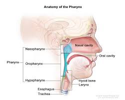
The pharynx is conventionally divided into three sections: the nasopharynx, oropharynx, and laryngopharynx.
The pharynx is for important in vocalization.
Two sets of pharyngeal muscles form the pharynx and determine the shape of its lumen: inner layer of longitudinal muscles and an outer circular layer.
The pharynx is innervated by the pharyngeal plexus of the vagus nerve.
Muscles in the pharynx push the food into the esophagus.
The pharynx joins the esophagus at the esophageal inlet which is located behind the cricoid cartilage.
The nasopharynx is the upper portion of the pharynx,and it extends from the base of the skull to the upper surface of the soft palate.
It includes the space between the internal nares and the soft palate and lies above the oral cavity.
The adenoids, also known as the pharyngeal tonsils, are lymphoid tissue structures located in the posterior wall of the nasopharynx.
Waldeyer’s tonsillar ring is an annular arrangement of lymphoid tissue in both the nasopharynx and oropharynx.
The nasopharynx is lined by respiratory epithelium that is pseudostratified, columnar, and ciliated.
Polyps or mucus can obstruct the nasopharynx, as can congestion due to an upper respiratory infection.
The eustacian tube, which connects the middle ear to the pharynx, opens into the nasopharynx at the pharyngeal opening of the auditory tube.
The opening and closing of the eustacian tubes serves to equalize the barometric pressure in the middle ear with that of the ambient atmosphere.
The anterior nasopharynx communicates through the choanae with the nasal cavities.
The lateral wall of the pharyngeal opening of the auditory tube, is a somewhat triangular shaped firm prominence, the torus tubarius or cushion, caused by the medial end of the cartilage of the tube that elevates the mucous membrane.
Two folds arise from the cartilaginous opening:
the salpingopharyngeal fold, a vertical fold of mucous membrane extending from the inferior part of the torus and containing the salpingopharyngeus muscle
the salpingopalatine fold, a smaller fold, in front of the salpingopharyngeal fold, extending from the superior part of the torus to the palate and containing the levator veli palatini muscle, and contains some muscle fibers called salpingopalatine muscle.
The tensor veli palatini is lateral to the levator and is deep to the cartilaginous opening.
The oropharynx lies behind the oral cavity, extending from the uvula to the level of the hyoid bone.
The oropharynx opens anteriorly, through the isthmus faucium, into the mouth, while in its lateral wall, between the palatoglossal arch and the palatopharyngeal arch, is the palatine tonsil.
The anterior wall consists of the base of the tongue and the epiglottic vallecula.
Its lateral wall is made up of the tonsil, tonsillar fossa, and tonsillar (faucial) pillars.
The superior wall consists of the inferior surface of the soft palate and the uvula.
The oropharynx is lined by non-keratinized squamous stratified epithelium.
Because both food and air pass through the pharynx, a flap of connective tissue called the epiglottis closes over the glottis when food is swallowed to prevent aspiration.
The HACEK organisms (Haemophilus, Actinobacillus actinomycetemcomitans, Cardiobacterium hominis, Eikenella corrodens, Kingella) are part of the normal oropharyngeal flora, which grow slowly, prefer a carbon dioxide-enriched atmosphere, and share an enhanced capacity to produce endocardial infections, especially in young children.
The laryngopharynx, also known as hypopharynx, is the caudal part of the pharynx.
it is the part of the throat that connects to the esophagus.
It lies inferior to the epiglottis and extends to the location where this common pathway diverges into the respiratory and esophageal pathways.
Like the oropharynx above it, the laryngopharynx serves as a passageway for food and air and is lined with a stratified squamous epithelium.
It is innervated by the pharyngeal plexus.
At that point, the laryngopharynx is continuous with the esophagus posteriorly.
The esophagus conducts food and fluids to the stomach; air enters the larynx anteriorly.
During swallowing, food passes, and the air passage temporarily stops.
The superior boundary of the laryngopharynx is at the level of the hyoid bone located between the 4th and 6th cervical vertebrae,
The laryngopharynx ( hypopharynx) includes three major sites: the pyriform sinus, postcricoid area, and the posterior pharyngeal wall.
The blood supply to the laryngopharynx includes the superior thyroid artery, the lingual artery and the ascending pharyngeal artery.
The neural supply to the hypopharynx is from both the vagus and glossopharyngeal nerves.
The vagus nerve provides an auricular branch which also supplies the external auditory canal, thus laryngopharyngeal cancer can result in referred ear pain.
The pharynx moves food from the mouth to the esophagus, moves air from the nasal and oral cavities to the larynx, is also used in speech, as pharyngeal consonants are articulated here.
Waldeyer’s tonsillar ring is an anatomical annular arrangement of lymphoid tissue in the pharynx.
Waldeyer’s ring circumscribes the naso- and oropharynx, with some of its tonsillar tissue located above and some below the soft palate.
The Waldeyer’s ring prevents the invasion of microorganisms from going into the air and food passages and this helps in the defense mechanism of the respiratory and alimentary systems.