 The parasympathetic nervous system (PSNS) is one of the two divisions of the autonomic nervous system, a division of the peripheral nervous system (PNS)), the other being the sympathetic nervous system.
The enteric nervous system (ENS) is has its own independent reflex activity.
The autonomic nervous system is responsible for regulating unconscious activities.
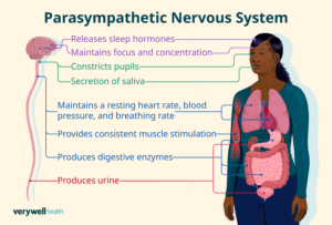
The PSNS is responsible for stimulation of “rest-and-digest” or “feed and breed”activities that occur when the body is at rest, especially after eating, including sexual arousal, salivation, lacrimation, urination, digestion and defecation.
The parasympathetic system functions include slowing of the heart, an increased rate of digestion, narrowing of the airways, promotion of urination, and sexual arousal.
The PSNS’s actions are complementary to that of the sympathetic nervous system, which is responsible for stimulating activities associated with the fight-or-flight response.
PSNS fibers arise from the central nervous system.
Specific parasympathetic nerves include several cranial nerves, specifically the oculomotor nerve, facial nerve, glossopharyngeal nerve, and vagus nerve.
Three spinal nerves in the sacrum (S2-4), commonly referred to as the pelvic splanchnic nerves, also act as parasympathetic nerves.
The parasympathetic nervous system is has a craniosacral outflow.
The sympathetic nervous system has a thoracolumbar outflow.
The parasympathetic nerves are autonomic or visceral branches of the peripheral nervous system (PNS).
Parasympathetic nerve supply arises through three primary areas:
cranial nerves in the cranium, namely the preganglionic parasympathetic nerves (CN III, CN VII, and CN IX) usually arise from specific nuclei in the central nervous system (CNS) and synapse at one of four parasympathetic ganglia: ciliary, pterygopalatine, otic, or submandibular.
From these four ganglia the parasympathetic nerves target tissues via trigeminal branches, the ophthalmic nerve, maxillary nerve, mandibular nerve.
The vagus nerve does not participate in these cranial ganglia.
Most vagal parasympathetic fibers are destined for ganglia on or near thoracic viscera- esophagus, trachea, heart, lungs, and abdominal viscera-stomach, pancreas, liver, kidneys, small intestine, and about half of the large intestine.
The vagus innervation ends between the midgut and hindgut, just before the splenic flexure of the transverse colon.
The pelvic splanchnic efferent preganglionic nerve cell bodies reside in the lateral gray horn of the spinal cord at the T12-L1 vertebral levels.
The conus medullaris axons exit the vertebral column as S2-S4 spinal nerves through the sacral foramina.
Axons of the PSNS continue away from the CNS to synapse at an autonomic ganglion.
The parasympathetic ganglion where these preganglionic neurons synapse will be close to the organ of innervation.
In the sympathetic nervous system, synapses between pre- and post-ganglionic efferent nerves in general occur at ganglia that are farther away from the target organ.
Efferent parasympathetic nerve signals are carried from the central nervous system to their targets by a system of two neurons.
The first neuron in this pathway is referred to as the preganglionic or presynaptic neuron.
The preganglionic cell body sits in the central nervous system and its axon usually extends to synapse with the dendrites of a postganglionic neuron somewhere else in the body.
The axons of presynaptic parasympathetic neurons are usually long.
The axons of presynaptic parasympathetic neurons extend from the CNS into a ganglion that is either very close to or embedded in their target organ.
The postsynaptic parasympathetic nerve fibers are very short.
The oculomotor nerve is responsible for parasympathetic functions related to the eye.
The oculomotor nerve parasympathetic fibers originate in the Edinger-Westphal nucleus in the central nervous system.
It travels through the superior orbital fissure to synapse in the ciliary ganglion located just behind the orbit.
From the ciliary ganglion the postganglionic parasympathetic fibers of the oculomotor nerve leave via short ciliary nerve fibers, a continuation of the nasociliary nerve (a branch of ophthalmic division of the trigeminal nerve (CN V1)).
The short ciliary nerves innervate the orbit to control the ciliary muscle, which is responsible for accommodation and the iris sphincter muscle, which is responsible for miosis or constriction of the pupil in response to light or accommodation.
The two aspects of the motor portion of the ocular nerve are known as somatic and visceral motor nerves.
The somatic motor is responsible for moving the eye in precise motions and for keeping the eye fixated on an object.
The visceral motor helps constrict the pupil.
The parasympathetic aspect of the facial nerve controls secretion of the sublingual and submandibular salivary glands, the lacrimal gland, and the glands associated with the nasal cavity.
The preganglionic fibers of the facial nerve originate within the CNS in the superior salivatory nucleus and leave as the intermediate nerve to connect with the facial nerve just distal to it surfacing the central nervous system.
Just after the facial nerve geniculate ganglion in the temporal bone, the facial nerve gives off two separate parasympathetic nerves: the greater petrosal nerve and the chorda tympani.
The greater petrosal nerve travels through the middle ear and
combines with the deep petrosal nerve to form the nerve of the pterygoid canal.
The parasympathetic fibers of the nerve of the pterygoid canal synapse at the pterygopalatine ganglion.
These parasympathetic fibers lead to the lacrimal gland and control tear production.
The pterygopalatine ganglion are the descending palatine nerves (CN V2 branch), which include the greater and lesser palatine nerves.
The greater palatine parasympathetic synapse on the hard palate and regulate mucus glands.
The lesser palatine nerve synapses at the soft palate and controls taste receptors and mucus glands.
Divisions from the pterygopalatine ganglion: posterior, superior, and inferior lateral nasal nerves; and the nasopalatine nerves are all branches of CN V2, maxillary division of the trigeminal nerve that bring parasympathetic innervation to glands of the nasal mucosa.
The second parasympathetic branch leaving the facial nerve is the chorda tympani, which carries secretomotor fibers to the submandibular and sublingual glands.
The chorda tympani travels through the middle ear and attaches to the lingual nerve the mandibular division of trigeminal, CN V3.
Preganglionic fibers synapse at the submandibular ganglion and send postganglionic fibers to the sublingual and submandibular salivary glands.
The glossopharyngeal nerve’s parasympathetic fibers innervate the parotid salivary gland.
The preganglionic fibers depart CN IX as the tympanic nerve and continue to the middle ear where they make up a tympanic plexus on the cochlear promontory.
The vagus nerve, has parasympathetic functions that originate in the dorsal nucleus of the vagus nerve and the nucleus ambiguus in the CNS.
The vagus nerve has an autonomic ganglion associated with it at approximately the level of C1 vertebra.
The vagus has no parasympathetic to the cranium.
The vagus nerve as it enters the thorax becomes the recurrent laryngeal nerve, which becomes the inferior laryngeal nerve.
The left vagus nerve the recurrent laryngeal nerve hooks around the aorta to travel back up to the larynx and proximal esophagus.
The right vagus nerve, the recurrent laryngeal nerve hooks around the right subclavian artery to travel back up to the same location as its counterpart.
Each recurrent laryngeal nerve supplies the trachea and the esophagus with parasympathetic secretomotor innervation for glands associated with them.
Nerves that comes off the vagus nerves approximately at the level of entering the thorax are the cardiac nerves.
Cardiac nerves go on to form cardiac and pulmonary plexuses around the heart and lungs.
The main vagus nerves continue into the thorax and links with the esophagus and sympathetic nerves from the sympathetic trunks to form the esophageal plexus.
The vagus nerve controls of the gut smooth muscles and glands.
The esophageal plexus enters the abdomen through the esophageal hiatus forming the anterior and posterior vagus trunks.
The vagus trunks join preaortic sympathetic ganglion around the aorta to disperse with the blood vessels throughout the abdomen.
Abdominal parasympathetic innervation occurs in the abdomen include the pancreas, kidneys, liver, gall bladder, stomach and intestines.
The vagus parasympathetic innervation continues until the end of the midgut, two thirds of the way across the transverse colon near the splenic flexure.
The pelvic splanchnic nervesS2-4, work in tandem to innervate the pelvic viscera.
Pelvic splanchnic nerves are composed of mixed autonomic nerve fibers, both parasympathetic and sympathetic, and include the vesical, prostatic, rectal, uterovaginal, and inferior hypogastric plexuses.
The preganglionic neurons in these pathway do not synapse in a ganglion as in the cranium.
These preganglionic neurons synapse
in the walls of the pelvic tissues or organs that they innervate.
The pelvic parasympathetic nerve pathway controls include those of the urinary bladder, ureters, urinary sphincter, anal sphincter, uterus, prostate, glands, vagina, and penis.
The parasympathetic nerves will unconsciously cause peristaltic movements of the ureters and intestines.
They move urine from the kidneys into the bladder and food down the intestinal tract and, will assist in excreting urine from the bladder or defecation.
Parasympathetic stimulation causes the bladder detrusor muscle to contract and simultaneously relaxes the internal sphincter muscle between the bladder and the urethra, allowing the bladder to void.
Similarly, parasympathetic stimulation of the internal anal sphincter relaxes this muscle to allow defecation.
Parasympathetic nerves plays a huge role in continence and bowel retention.
The afferent fibers of the autonomic nervous system of organs of the body, are not divided into parasympathetic and sympathetic fibers as the efferent fibers are.
General visceral afferent sensations are mostly unconscious visceral motor reflex sensations from hollow organs and glands that are transmitted to the CNS.
The heart’s sinoatrial pacing contributes to maintain the heart rate in the range of 60-100 beats per minute.
The two branches of the autonomic nervous system act in a complementary way increasing or slowing the heart rate.
The vagus nerve acts on sinoatrial node slowing its conduction thus actively modulating vagal tone.
The vagal nerve modulation is mediated by the neurotransmitter acetylcholine and causes downstream changes to ionic currents and calcium of heart cells.
Increased vagal tone is associated with a diminished and more variable heart rate.
The parasympathetic nervous system acts on vascular and cardiac control via respiratory sinus arrhythmia: rhythmical fluctuation of heart rate at the respiration frequency, characterized by heart rate increase during inspiration and decrease during expiration.
Parasympathetic activity on cavernous nerves from the prostatic plexus stimulate smooth muscles in the fibrous trabeculae of the arteries of penis to relax and allow blood to fill the two corpora cavernosa and the corpus spongiosum of the penis, making it rigid to prepare for sexual activity.
Upon emission of ejaculate, the sympathetics participate and cause peristalsis of the ductus deferens and closure of the internal urethral sphincter to prevent semen from entering the bladder.
Parasympathetics fibers cause peristalsis of the urethral muscle, and the pudendal nerve causes contraction of the bulbospongiosus, to forcibly emit the semen.
During remission the penis becomes flaccid again.
In the female, the erectile tissue analogous to the male, yet less substantial, and plays a large role in sexual stimulation.
The PN system fibers cause release of secretions in the female that decrease friction.
The parasympathetic nervous system innervates the fallopian tubes, which helps peristaltic contractions and movement of the oocyte to the uterus for implantation, and aid in secretions from the female genital tract promoting sperm migration.
The parasympathetic nervous system uses chiefly acetylcholine.
Acetylcholine acts on two types of receptors, the muscarinic and nicotinic cholinergic receptors.
Acetylcholine transmissions occur in two stages: When stimulated, the preganglionic neuron releases ACh at the ganglion, which acts on nicotinic receptors of postganglionic neurons.
The postganglionic neuron then releases ACh to stimulate the muscarinic receptors of the target organ.
The five main types of muscarinic receptors:
The M1 muscarinic receptors are located in the neural system.
The M2 muscarinic receptors are located in the heart, and act to bring the heart back to normal after the actions of the sympathetic nervous system.
M2 muscarinic receptors slow down the heart rate, reducing contractile forces of the atrial cardiac muscle, and reducing conduction velocity of the sinoatrial node and atrioventricular node.
M2 muscarinic receptors have a minimal effect on the contractile forces of the ventricular muscle due to sparse innervation of the ventricles from the parasympathetic nervous system.
The M3 muscarinic receptors (CHRM3) are located at many places in the body, such as the endothelial cells of blood vessels, as well as the lungs causing bronchoconstriction. The net effect of innervated M3 receptors on blood vessels is vasodilation, as acetylcholine causes endothelial cells to produce nitric oxide, which diffuses to smooth muscle and results in vasodilation. They are also in the smooth muscles of the gastrointestinal tract, which help in increasing intestinal motility and dilating sphincters. The M3 receptors are also located in many glands that help to stimulate secretion in salivary glands and other glands of the body. They are also located on the detrusor muscle and urothelium of the bladder, causing contraction.
The M4 muscarinic receptors: Postganglionic cholinergic nerves, possible CNS effects
The M5 muscarinic receptors: Possible effects on the CNS
Types of nicotinic receptors
In vertebrates, nicotinic receptors are broadly classified into two subtypes based on their primary sites of expression: muscle-type nicotinic receptors (N1) primarily for somatic motor neurons; and neuronal-type nicotinic receptors (N2) primarily for autonomic nervous system.[
Relationship to sympathetic nervous system
Sympathetic and parasympathetic divisions typically function in opposition to each other. The sympathetic division typically functions in actions requiring quick responses. The parasympathetic division functions with actions that do not require immediate reaction. A useful mnemonic to summarize the functions of the parasympathetic nervous system is SSLUDD (sexual arousal, salivation, lacrimation, urination, digestion and defecation).
The functions promoted by activity in the parasympathetic nervous system are associated with our day-to-day living. The parasympathetic nervous system promotes digestion and the synthesis of glycogen, and allows for normal function and behavior.
Parasympathetic action helps in digestion and absorption of food by increasing the activity of the intestinal musculature, increasing gastric secretion, and relaxing the pyloric sphincter.
The parasympathetic nervous system is called the rest and digest division of the autonomic nervous system.