 Paranasal sinuses are a group of four paired air-filled spaces that surround the nasal cavity.
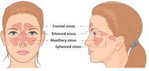
The maxillary sinuses are located under the eyes; the frontal sinuses are above the eyes; the ethmoidal sinuses are between the eyes and the sphenoidal sinuses are behind the eyes.
The sinuses are named for the facial bones in which they are located.
All the Paranasal sinuses are innervated by branches of the trigeminal nerve (CN V).
The maxillary sinuses are the largest of the paranasal sinuses, and are under the eyes, in the maxillary bones.
The maxillary sinuses open in the back of the semilunar hiatus of the nose, and are innervated by the maxillary nerve (CN V5).
The frontal sinuses, superior to the eyes, in the frontal bone, which forms the hard part of the forehead, and are innervated by the ophthalmic nerve (CN V2).
The ethmoidal sinuses, which are formed from several discrete air cells within the ethmoid bone between the nose and the eyes.
They are innervated by the ethmoidal nerves, which branch from the nasociliary nerve of the ophthalmic nerve (CN V2).
The sphenoidal sinuses, in the sphenoid bone are innervated by the ophthalmic and maxillary nerve (CN V2and V5).
The paranasal sinuses are lined with ciliated pseudostratified columnar epithelium.
A function of the paranasal sinuses is the production of nitric oxide, which also functions as a facilitator of oxygen uptake.
The bones of the facial skull are the attachment points for the facial muscles.
The bones of the facial skull is important because the bones are the attachment points for the facial muscles, and provide some resistance in case of injury.
The facial bones also Isolate sensitive structures: roots of teeth, eyeballs from rapid temperature fluctuations in the nasal cavity during inhalation and exhalation:
Humidification and warming of inhaled air occurs a result of slow airflow in the sinuses.
The paranasal sinus acts as a sensory system of air signals, a baroreceptor organ, that responds to changes in environmental pressure.
The paranasal sinuses form developmentally through excavation of bone by air-filled sacs from the nasal cavity, a process begins prenatally
and it continues through the course of a lifetime.
The natural ventilation rate of a sinus with a single sinus ostium is extremely slow, and such limited ventilation may be protective for the sinus to help prevent drying of its mucosal surface and maintain a near-sterile environment with high carbon dioxide concentrations and minimal pathogen access.
The gas content of the maxillary sinus is similar to venous blood, with high carbon dioxide and lower oxygen levels compared to breathing air.
At birth, only the maxillary sinus and the ethmoid sinus are developed but not yet pneumatized: by the age of seven they are fully aerated.
The sphenoid sinus appears at the age of three, and the frontal sinuses first appear at the age of six, and fully develop during adulthood.
Coronal CT scan of the paranasal sinuses (Bone)
The paranasal sinuses are joined to the nasal cavity via small orifices called ostia.
Ostia are blocked easily by allergic inflammation, or by swelling in the nasal lining that occurs with a cold, impairing normal drainage of mucus within the sinuses and sinusitis may occur.
The maxillary posterior teeth are close to the maxillary sinus, and the presence are of an infection can involve these teeth.
Inflammation of the sinuses can occur with infection of the adjacent teeth.
Sinus inflammation may be treated with drugs such as decongestants, which cause vasoconstriction in the sinuses; reducing inflammation by traditional nasal irrigation; or by corticosteroids.
Malignancies of the paranasal sinuses comprise approximately 0.2% of all malignancies.
About 80% of these malignancies arise in the maxillary sinus.
Men are much more often affected than women.
Paranasal cancers most often occur in the age group between 40 and 70 years.
Carcinomas are more frequent than sarcomas.
Metastases are rare.
Tumors of the sphenoid and frontal sinuses are extremely rare.