 Cornerstone of treatment for symptomatic bradyarrhythmias.
Pacemakers provide an artificial electrical stimulus to initiate a cardiac cycle.
Indications for permanent pacemaker implantation include sinus node dysfunction with documented symptomatic bradycardia, symptomatic chronotropic incompetence, and symptomatic sinus bradycardia that results from required drug therapy for medical disorders.
Estimated 200,000 placed each year in the US and more than 5 million have pacemakers.
The system consists of a pulse generator and 1, 2 or three leads.
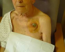
The pulse generator, is generally implanted under the skin just below the collarbone on either the right or left side.
The generator contains the battery and electrical circuitry and is connected to the leads and is implanted in the chest pectoral region.
The leads are connected to the right atrium, the right ventricle or the coronary sinus (the left ventricle).
The leads are inserted using x-ray control, via a vein found in this area, and positioned in the appropriate right-sided heart chamber.
The leads are tested before the pulse generator is attached and the incision is closed.
Pulse generator emits electrical pulses that depolarize the myocardium.
Sense electrical signals from cardiac chambers and respond by inhibition of pacing or tracking the senses event with a pacing pulse.
The pacemaker mode refers to the chamber to be paced-atrium, ventricle or dual, the chambers to be sensed-atrium, ventricle or dual, the response to sensing-inhibit, trigger or dual, the presence or absence of rate modulation and multisite pacing.
Single chamber atrial or ventricular pacemakers sense signal from corresponding cardiac chambers and deliver a pacing stimulus if no signal is sensed at he programmed rate.
Dual-chamber pacemakers sense and pace both the atrium and the ventricle.
Most dual chambered pacemakers are able to switch between different pacing modes as activated by changes in the atria rhythm that is present.
For patients with sick sinus syndrome, dual chamber pacing has been shown to improve outcomes.
Single chamber, ventricular pacing and dual chamber pacing have a similar effect on outcomes in patients with high-grade AV block, dual chamber pacing is preferred for most patients in order to prevent the pacemaker syndrome.
Rate adaptive pacemakers are helpful when blunted heart rates occur in response to exercise.
With rate adaptive pacemakers special sensors are present that, when activated by monitoring body movements during exercise, increase the pacing rate.
May contain sensors that can detect changes in minute ventilation by measuring thoracic impedance with ventilation changes or sensors that can detect changes in the QT interval reflecting sympathetic drive.
May contain a combination of sensors to achieve optimal results.
Cornerstone of treatment for symptomatic bradyarrhythmias.
AV dissociation or retrograde atrial activation may occur resulting in a pacemaker syndrome.
About 10% of patients who have bradycardia pacing have complications within five years after implantation of a conventional pacemaker.
To avoid pacemaker pocket and lead related complications, leadless pacemakers have been developed:risk of complications with leadless pacemakers is 31 to 63% lower than the risk with conventional intravenous pacemakers during the first year after implantation.
Implantation of the leadless pacemaker is associated with a higher risk of cardiac perforation, with a 0.4 percentage increase over conventional pacemakers.
Leadless pacemakers are associated with a much lower infection rate.
Pacemaker syndrome may occur in up to 30% of patients, and is associated with hemodynamic, neurohormonal and autonomic changes.
Pacemaker syndrome leads to disabling symptoms with dizziness, weakness, heart failure and syncope.
The pacemaker syndrome can lead to the development of atrial fibrillation and increased risk of stroke.
Pacemaker syndrome can be prevented or reversed by restoring AV synchrony.
Long-term right ventricular apical pacing causes pathological changes and increased morbidity.
Right ventricular pacing may induce cardiomyopathy which ranges from 6 to 25%.
Pacing induced cardiomyopathy occurs when the left ventricular ejection fraction falls to 50% or less coupled with an absolute reduction of 5 to 10 percentage points from the baseline left ventricular ejection fraction.
Risk factors for pacing induced cardiomyopathy include older age, male sex, history of atrial fibrillation, a wider paced QRS, duration, left ventricular dysfunction at baseline, and a high burden of right ventricular pacing.
With a right ventricular pacing burden that exceeds 20%, cardiac physiologic pacing has become standard to reduce the risk of cardiomyopathy:cardiac physiologic pacing can be achieved through resynchronization therapy involving biventricular, pacing or with conduction system. pacing, restoring, or preserving, the synchrony of ventricular contraction.
Ventricular dyssynchrony that arises from right ventricular pacing can cause cardiac dysfunction and heart failure.
If ventricular pacing occurs without atrial synchronization a loss in the atrial contribution to ventricular filling occurs.
Biventricular pacing has improved the treatment and outcomes of heart failure and the reduced ejection fraction.
Indications for a biventricular pacing include a left ventricular ejection fraction of 35% or lower, left bundle branch block, a QRS duration of 150 ms or longer and a class III or ambulatory class IV NYHA.despite optical medical therapy for heart failure,
Benefits of biventricular pacing are determined by patient characteristics, the location of the left ventricular lead to non-apical lateral or posterior lateral locations and optimal programming of device to ensure more than 97% by ventricular pacing.
Factors that portend, a higher likelihood of benefit from biventricular pacing include: left bundle, branch block, nonischemic cardiomyopathy, and female gender.
Women derive more benefit from by ventricular duration at a shorter QRS duration.
Dual chamber pacing provides hemodynamic advantages by coordinating the timing of atrial and ventricular systole.
Dual chamber promotes physiological heart rate response inpatients within sinus node function and AV block.
Paced rhythms can mask the diagnosis of acute myocardial infarction by interfering with the recognition of new Q waves, ST-segment changes, and T-wave inversions.
Able to store information useful in clinical management, follow-up and trouble shooting: reveals pacing mode, pacing rates, battery information, lead information, heart rate histograms, event counters, pacing frequency, arrhythmias, mode switches, activity levels, fluid status, monitoring of threshold and sensing tests and ability to correlate symptoms with atrial or ventricular arrhythmias.
Goal is to mimic normal electrical activation of the heart.
The rate of stroke is high in patients who have a pacemaker, with stroke occurring in 5.8% of patients within four years of implantation (Healey JS et al).
Estimated that between 50% and 75% of patients with pacemakers will require an MRI over the lifetime of the device.
Between 14 and 35% will require long-term oral anticoagulation therapy providing a peri-procedural treatment dilemma for implantation.
A person with an artificial cardiac pacemaker will require one or two pacemaker checks per year.
Adjustments to the pacemaker can be made non-invasively using a specially designed radio frequency programmer, with a wand placed on the skin over the implanted device.
Most pacemakers last longer than five years.
The life of the battery is dependent on how it’s used.
Most pacemakers can calculate the remaining life of the battery and this information is available.
Because transvenous pacemakers are subject to lead in pocket related complications self-contained, leadless, pacemakers are designed to be placed in the right ventricle to mitigate these complications.
Single chamber ventricular pacemakers do not provide atrial pacing or consistent atrioventricular synchrony, thus limiting leadless pacemaker therapy to approximately 20% of patients who have an indication for a pacemaker.
A dual chamber leadless pacemaker system that can provide atrial pacing and reliable atrial ventricular synchrony is under development.
A pacemaker in wireless communication with a subcutaneous implantable cardio converter-defibrillator (ICD) exceeded performance goals for freedom from major complications related to the leadless pacemaker, for communication between the leadless pacemaker and subcutaneous ICD and for the percentage of patients with a appropriate pacing threshold pulse with at six months(MODULAR ATP investigators).