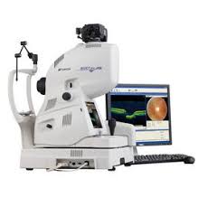
Optical coherence tomography (OCT) is an imaging technique that uses low-coherence light to capture micrometer-resolution.
It allows for two- and three-dimensional imaging.
OCT uses a low coherence light source that illuminates an interferometer at its core to detect the interference between reflected light from scatterers at multiple depths within tissue, and the reference reflector in order to extract information about their depth location and reflectivity.
It allows segmental visualization of retinal and choroidal features and detailed rapid examination of neurodegenerative and neurovascular components of age related macular degeneration.
OCT is sensitive to low light levels of returned light and reduces exposure of sensitive tissues to light of high intensity.
OCT has a wide range of differentiation in the gradations of reflectivity of internal tissues, with resolution in several microns, allowing visualization of cellular and subcellular layers at a depth of a millimeter in most tissues.
Its use of light in the near infrared range is comfortable for patients, unlike white light from ophthalmoscopes.
It is used in diagnosing age related macular degeneration and evaluating the stage, assessing disease progression, guiding treatment decisions and assessing response to treatment.
Provides high resolution diagnostic visualization of ocular tissues: cornel, lens, and retina and can be used to obtain images of vascular, endothelial structures, and atherosclerosis.
It enables optical biopsy: imaging subsurface, microscopic tissue structures, in situ, and in real time, without the need to biopsy specimens in a conventional way for histopathology.
It is used for medical imaging.
It typically employs near-infrared light wavelength, which allows it to penetrate into the scattering medium.
Light source: superluminescent diodes, ultrashort pulsed lasers, and supercontinuum lasers, allow optical coherence tomography sub-micrometer resolution with very wide-spectrum sources emitting over a ~100 nm wavelength range.
OCT systems are employed in art conservation and diagnostic medicine, notably in ophthalmology and optometry where it can be used to obtain detailed images from within the retina.
In OCT, interference is shortened to a distance of micrometers, owing to the use of broad-bandwidth light sources.
OCT provides tissue morphology imagery at much higher resolution (less than 10 μm axially and less than 20 μm laterally, than other imaging modalities such as MRI or ultrasound.
Its benefits are:
Live sub-surface images at near-microscopic resolution
Instant, direct imaging of tissue morphology
No preparation of the sample or subject, no contact
No ionizing radiation
OCT delivers a high resolution because it is based on a light optical beam directed at the tissue, and a small portion of this light that reflects from sub-surface features is collected.
Most light is not reflected but, rather, scatters off at large angles.
OCT is limited to imaging 1 to 2 mm below the surface in biological tissue.
No special preparation of a biological specimen is required, and the laser output from the instruments is low using eye-safe near-infrared light is used and no damage to the sample is expected.
OCT is used by ophthalmologists and optometrists to obtain high-resolution images of the retina and anterior segment.
OCT provides a straightforward method of assessing cellular organization, photoreceptor integrity, and axonal thickness in glaucoma, macular degeneration, diabetic macular edema,multiple sclerosis and other eye diseases or systemic pathologies which have ocular signs.
Ocular OCT is used heavily by ophthalmologists and optometrists to obtain high-resolution images of the retina and anterior segment.
Owing to OCT’s capability to show cross-sections of tissue layers with micrometer resolution, OCT provides a straightforward method of assessing cellular organization, photoreceptor integrity, and axonal thickness in glaucoma, macular degeneration, diabetic macular edema, multiple sclerosis and other eye diseases or systemic pathologies which have ocular signs.
OCT can assess the vascular health of the retina via a OCT angiography.
OCT is used to image coronary arteries in order to visualize vessel wall lumen morphology and microstructure at a resolution 10 times higher than other existing modalities such as intravascular ultrasounds and x-ray angiography.
Intravascular OCT has been investigated for use in coronary arteries, and endovascular treatment of ischemic stroke and brain aneurysms.
Endoscopic OCT has been applied to the detection and diagnosis of cancer and precancerous lesions, such as Barrett’s esophagus and esophageal dysplasia.
OCT has been applied to the diagnosis of various skin lesions including carcinomas.
Optical coherence tomography (OCT) is a noninvasive tool for a basal cell carcinoma diagnosis which can generate real time, cross-sectional imaging of tissue micro architecture with a depth of 1 to 1.5 mm: it is not inferior to regular care punch biopsy.
OCT can detect enamel white spot lesions around and beneath the orthodontic brackets.
Fiber-based OCT systems are adaptable to industrial environments.
