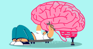
Neuroplasticity refers to the brain’s ability to change and adapt throughout a person’s life.
Neuroplasticity, also known as neural plasticity, or brain plasticity, is the ability of neural networks in the brain to change through growth and reorganization.
The brain has the ability to reorganize itself in response to learning, experience, and even injury.
Neuroplasticity allows the brain to form new neural connections, strengthen existing ones, and even reassign certain functions from damaged areas to undamaged ones.
Neuroplastic adaptability plays a role in the ability to learn new things, recover from injuries or diseases that affect the brain, and adapt to changes in our environment.
Engaging in new experiences, learning new skills, and maintaining an active and stimulating lifestyle can promote neuroplasticity and help keep the brain healthy and functioning optimally.
Neuroplasticity is when the brain is rewired to function in some way that differs from how it previously functioned.
Changes range from individual neuron pathways making new connections, to systematic adjustments like cortical remapping or neural oscillation.
Neuroplasticities include circuit and network changes that result from learning a new ability, information acquisition, environmental influences, practice, and psychological stress.
Neuroplasticity’s many aspects of the brain can be altered even through adulthood, however, the developing brain exhibits a higher degree of plasticity.
Activity-dependent plasticity can have significant implications for healthy development, learning, memory, and recovery from brain damage.
The term neuronal plasticity is used to describe nonpathological changes in the structure of adult brains.
In people recovering from stroke neuroplasticity can occur as regions of the brain that remained healthy could sometimes take over, at least in part, functions that had been destroyed.
Neuroscientists suggest that brain exercises may be as useful as drugs to treat diseases as severe as schizophrenia.
Plasticity exists throughout life and that radical improvements in cognitive functioning –learning, think, perceiving, and remembering are possible even in the elderly.
Neuroplastic pathways are mainly signaling cascades, allow for gene expression alterations that lead to neuronal changes, and thus neuroplasticity.
Other factors play a role in the biological processes underlying the changing of neural networks in the brain, including synapse regulation via phosphorylation, the role of inflammation and inflammatory cytokines, proteins such as Bcl-2 proteins and neutrophorins, and energy production via mitochondria.
Neuroplasticity refers to the ability to make adaptive changes related to the structure and function of the nervous system.
There are two types of neuroplasticity: structural neuroplasticity and functional neuroplasticity.
Structural plasticity relates to the brain’s ability to change its neuronal connections: New neurons are constantly produced and integrated into the central nervous system.
Multiple cross-sectional imaging methods MRI), and computerized tomography (CT)) are used to study the structural alterations of the human brains.
The changes of grey matter proportion or the synaptic strength in the brain are related structural neuroplasticity.
Functional plasticity refers to the brain’s ability to alter and adapt the functional properties of neurons.
Functional plasticity: four known ways namely homologous area adaptation, map expansion, cross- model reassignment, and compensatory masquerade.
Homologous area adaptation of a cognitive task is shifted from a damaged part of the brain to its homologous area in the brain.
Map expansion relates to particular cognitive tasks that expand due to frequent exposure to stimuli.
Cross- model reassignment involves reception of novel input signals to a brain region which has been stripped off its input.
The changes can occur in response to previous activity to acquire memory or in response to malfunction or damage of neurons to compensate a pathological event.
Functions from one part of the brain may be transferred to another part of the brain based on the demand to produce recovery of behavioral or physiological processes.
Regarding physiological forms of activity-dependent plasticity, those involving synapses are referred to as synaptic plasticity.
The strengthening or weakening of synapses that results in an increase or decrease of firing rate of the neurons are called long-term potentiation (LTP) and long-term depression (LTD), respectively, and examples of synaptic plasticity that are associated with memory.
The cerebellum is a typical structure with redundancy within the circuitry, allowing plasticity at several sites.
Synaptic plasticity can be complemented by dependent plasticity involving the intrinsic excitability of neurons, which is referred to as intrinsic plasticity, where training alters the strength of functional connections contributing to encoding memories.
Cortical and subcortical rewiring of neuronal circuits can occur in response to training as well as in response to injury.
Neuroplasticity brain activity is associated with a given function can be transferred to a different location.
This activity can result from normal experience and also occurs in the process of recovery from brain injury.
Neuroplasticity explains improvements in functional outcomes with physical therapy post-stroke when rehabilitation techniques that support cortical reorganization as the mechanism of change include constraint-induced movement therapy, functional electrical stimulation, treadmill training with body-weight support, and virtual reality therapy.
Humans are thought had to acquire binocular vision, in particular stereopsis, in early childhood but recent years improvement in persons with amblyopia, convergence insufficiency or other stereo vision anomalies are examples of neuroplasticity have been demonstrated.
In the phenomenon of phantom limb sensation is based on the concept of neuroplasticity, as a person continues to feel pain or sensation within a part of their body that has been amputated, and it occurs in 60–80% of amputees: the cortical maps of the removed limbs are believed to have become engaged with the area around them in the postcentral gyrus.
The chronic pain experience of prolonged pain at sites that may have been previously injured, yet are otherwise currently well is related to neuroplasticity due to a maladaptive reorganization of the nervous system, both peripherally and centrally.
During the period of tissue injury, noxious stimuli and inflammation from the damage cause an increase in nociceptive input from the periphery to the central nervous system.
The prolonged nociception input from the periphery elicits a neuroplastic response at the cortical level to change its somatotopic organization for the painful site, inducing central sensitization.
Chronic pain significantly reduce the volume of grey matter in the brain globally, and more specifically at the prefrontal cortex and right thalamus.
Following treatment, these abnormalities in cortical reorganization and grey matter volume are resolved, as well as their symptoms: Similar findings reported for phantom limb pain, chronic low back pain and carpal tunnel syndrome.
Meditation may lead to change in the physical structure of brain regions associated with attention, anxiety, depression, fear, anger, and compassion as well as the ability of the body to heal itself.
Artistic engagement can create changes in neural network connections as well as increase cognitive flexibility, as demonstrated that long-term, artistic training can imprint a neural network system of spontaneous activity in which the related brain regions become functionally and topologically modularized in specific manners.
When the brain is repeatedly exposed to artistic training over long periods develop adaptations to make such activity both easier and more likely to spontaneously occur: neuroplasticity.
Aerobic exercise increases the production of neurotrophic factors that promote growth or survival of neurons, such as brain-derived neurotrophic factor (BDNF), insulin-like growth factor 1 (IGF-1), and vascular endothelial growth factor (VEGF).
Exercise-induce changes on the hippocampus that are associated with measurable improvements in spatial memory.
Consistent aerobic exercise over a period of several months induces marked clinically significant improvements in executive function, the cognitive control of behavior, and increases gray matter volume in multiple brain regions, particularly those that give rise to cognitive control.
The brain structures with the greatest improvements in gray matter volume in response to aerobic exercise: prefrontal cortex and hippocampus; moderate improvements are seen in the anterior cingulate cortex, parietal cortex, cerebellum, caudate nucleus, and nucleus accumbens.
Higher physical fitness scores as measured by VO2 max, are associated with better executive function, faster processing speed, and greater volume of the hippocampus, caudate nucleus, and nucleus accumbens.
With hearing loss in deaf and/or hard of hearing people the auditory cortex and other association areas of the brain undergo compensatory plasticity, to serve other functions, especially for vision and somatosensation.
The deaf have enhanced peripheral visual attention, better motion change detection ability in visual tasks, more effective visual search, and faster response time for visual targets compared to hearing individuals: this is neuroplasticity.
In deaf people, often times there is repurposing of other brain areas including primary auditory cortex, posterior parietal association cortex, and anterior cingulate cortex.
Altered visual processing in deaf people is often found to be associated with the repurposing of other brain areas including primary auditory cortex, posterior parietal association cortex (PPAC), and anterior cingulate cortex.
Brain areas that serve a function in auditory processing repurpose to process somatosensory information in congenitally deaf people.
The deaf have higher sensitivity in detecting frequency change in vibration above threshold and higher and more widespread activation in auditory cortex under somatosensory stimulation.
Neuroplasticity is involved in the development of sensory function.
The immature brain at birth adapts to sensory inputs after birth.
Congenital hearing loss, a rather frequent inborn condition affecting 1 of 1000 newborns, affects auditory development, and implantation of a sensory prostheses activating the auditory system has prevented the deficits and induced functional maturation of the auditory system.
There is also a sensitive period for such intervention within the first 2–4 years of life.
In prelingually deaf children, early cochlear implantation allows the children to learn the mother language and acquire acoustic communication.
The visual cortex in blind people may undergo cross-modal plasticity, and therefore other senses may have enhanced abilities.
Human echolocation is a learned ability to sense one’s environment from echoes.
This ability is used by some blind people to navigate their environment and sense their surroundings in detail.
Parts of the brain associated with visual processing are adapted for the skill of echolocation.
Individuals with attention deficit hyperactivity disorder (ADHD) have smaller volumes of the nucleus accumbens, amygdala, caudate, hippocampus, putamen, and overall cortical and intracranial volume, and have less surface area and cortical thickness, compared to people without ADHD.
Brain volume does not correlate to intelligence, or intelligence quotient (IQ).
People with ADHD exhibit atypical neuroconnectivity.
ADHD symptomatology may arise from a deviation from neurotypical synchronization and interaction within and between large-scale networks during brain development.
MRI and electroencephalography (EEG) studies in ADHD suggest that the long-term treatment of ADHD with stimulants, such as amphetamine or methylphenidate, decreases abnormalities in brain structure and function and improves function in several parts of the brain, such as the right caudate nucleus of the basal ganglia, left ventrolateral prefrontal cortex (VLPFC), and superior temporal gyrus.
Neuroplasticity is most active in childhood as a part of normal human development.
Trauma negatively affects many areas of the brain and puts a strain on the sympathetic nervous system from constant activation.
Trauma thus alters the brain’s connections, and children who have experienced trauma may be hyper vigilant or overly aroused.
Such adverse effects can be altered by actions of neuroplasticity.
Neuroplasticity has four different categories in children: impaired, excessive, adaptive, and plasticity.
Musical training can contribute to experience dependent structural plasticity.
Neuroplastic changes in the brain occur based on experiences that are unique to an individual, and can be a form of intervention for children with developmental disorders and neurological diseases.
Genes that play central roles in synaptic plasticity are the most significantly affected by age, showing reduced expression over time.
Reactive oxygen species appear to have a significant role in the regulation of synaptic plasticity and cognitive function.
Age-related increases in reactive oxygen species may also lead to impairments in these functions.
People who study more than one language have better cognitive functions and flexibilities than people who only speak one language.
Bilinguals are found to have longer attention spans, stronger organization and analyzation skills, and a better theory of mind than monolinguals.
The effect of multilingualism on better cognition is due to neuroplasticity.
Grey-matter density in the inferior parietal cortex for multilinguals were significantly greater than monolinguals.
Learning multiple languages not only re-structures the brain but also boosts brain’s capacity for plasticity.
There is increased myelinations in white matter tracts in bilingual individuals who actively used both languages in everyday life.
Using more than one language requires more efficient connectivity within the brain, which resulted in greater white matter density for multilinguals.
It is suggested that environmental, social experience in early multilinguals affects the structural and functional reorganization in the brain.
The elevation of norepinephrine, serotonin, and dopamine elicited by traditional antidepressants is quick, yet there is a significant delay in clinical efficacy and often inadequate treatment response.
There is an inverse relationship with the number of synapses and severity of depression symptoms and discovered that traditional antidepressants improved neuroplasticity but over a significantly protracted time course of weeks or months.
Ketamine, has potent anti-depressant effects after a single infusion due to its capacity to rapidly increase the number of dendritic spines and to restore aspects of functional